
Chordae Tendineae Collection
"Chordae Tendineae: The Vital Heart Strings that Keep the Rhythm in Harmony" In the intricate world of heart chambers and sinus node

All Professionally Made to Order for Quick Shipping
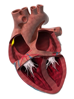
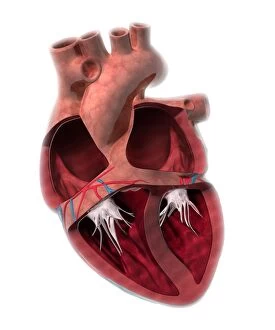
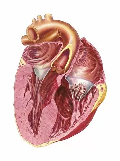
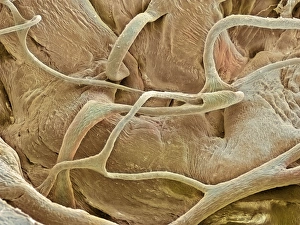
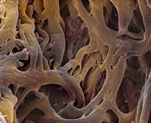
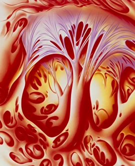
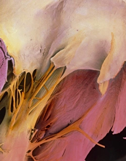
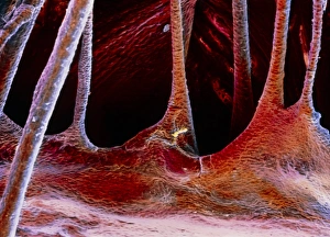
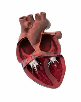
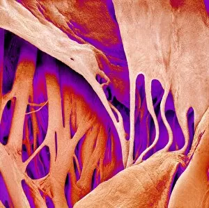
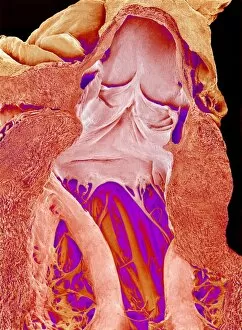
"Chordae Tendineae: The Vital Heart Strings that Keep the Rhythm in Harmony" In the intricate world of heart chambers and sinus node, chordae tendineae play a crucial role. These delicate tendons, depicted beautifully in artwork C014 / 2030 and C014 / 2029, connect the heart chamber walls to its valves. They act as anchors, preventing valve flaps from reversing into the wrong chamber during each heartbeat. However, sometimes these vital strings face challenges. Heart chamber wall defects can disrupt their function and lead to complications. Studying them under a scanning electron microscope (SEM) reveals their fascinating structure - thin yet resilient fibers that ensure proper valve closure. The mitral valve of a human heart comes alive through a captivating colored SEM image. Its chordae tendineae stand out like vibrant threads holding everything together with precision. Another SEM snapshot showcases a portion of a cardiac valve in false color, highlighting the complexity hidden within. An inside view of the right ventricle captured in an artwork provides insight into how chordae tendineae integrate seamlessly with other components of internal heart anatomy. Their presence is undeniable; they are truly indispensable for maintaining cardiac health. Multiple SEM images emphasize just how essential these tiny but mighty structures are - "Heart valve and strings, " they whisper repeatedly through detailed visuals.