
Cardiology Collection
Cardiology, the captivating realm of the heart, unveils its intricate beauty through a myriad of hints


All Professionally Made to Order for Quick Shipping
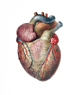
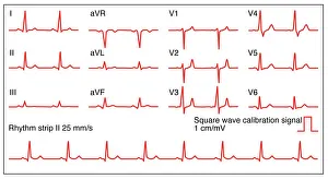
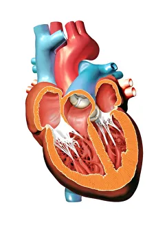
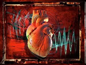
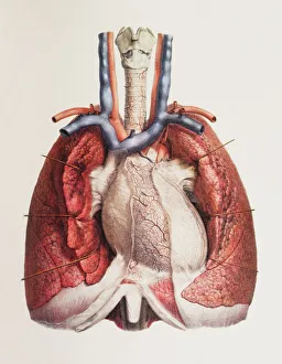
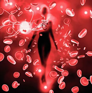
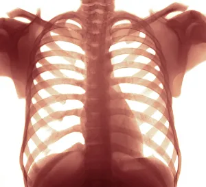
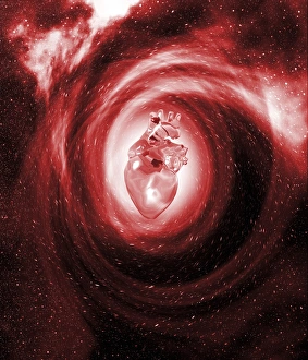
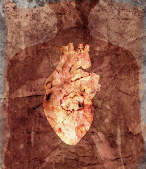
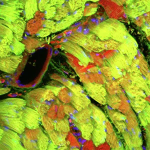
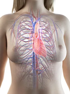
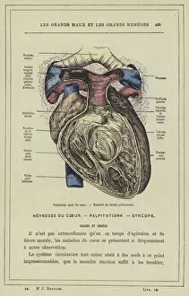
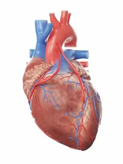
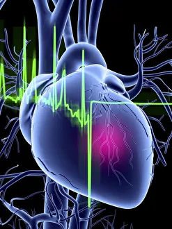
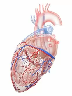
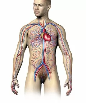
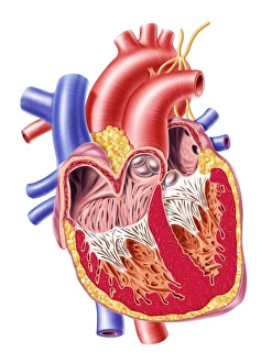
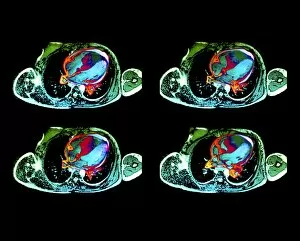
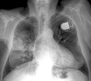
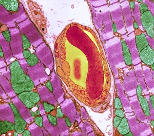
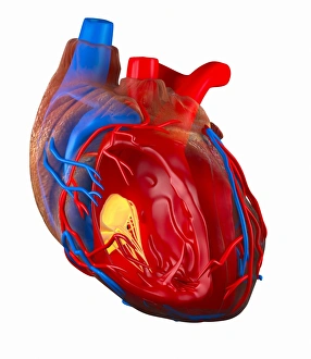
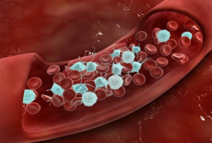
Cardiology, the captivating realm of the heart, unveils its intricate beauty through a myriad of hints. From ECGs showcasing the rhythmic dance of a normal heart rate to mesmerizing artwork depicting the human heart's anatomy, this field unravels the secrets held within our most vital organ. In awe-inspiring illustrations, artists bring to life the complexity and elegance of the human heart. Their brushstrokes breathe life into each chamber and vessel, while their colors mirror its vibrant energy. The harmonious connection between our hearts and lungs is depicted in breathtaking images that celebrate their symbiotic relationship. A chest X-ray reveals a healthy human heart, beating with strength and vigor—a testament to its resilience. Even in space, where gravity loses its grip on earthly matters, the heart remains an enigma; an ethereal image floating amidst celestial wonders. Resin casts capture every delicate detail of intricate blood vessels that intricately weave throughout this remarkable organ. These lifelines carry crimson red blood cells on their journey through arteries and veins—nourishing every corner of our being. Yet not all hearts are untouched by affliction; some bear witness to disease's cruel touch. In haunting depictions, we confront these realities head-on—the weakened muscle struggling against adversity—an urgent reminder for us to cherish our own health. Delving into history's annals takes us back centuries ago when anatomists meticulously studied hearts with fervor unmatched. Their discoveries paved pathways towards understanding cardiac physiology—a legacy still cherished today. Cardiology encompasses more than just medical expertise—it intertwines artistry with science as it seeks answers about one of humanity's greatest mysteries: our own heartbeat. It reminds us that within each pulsating rhythm lies a story waiting to be told—a tale written by countless generations who have marveled at this wondrous symbol of life itself.