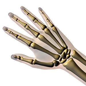
Hand bones and ligaments

![]()
Wall Art and Photo Gifts from Science Photo Library
Hand bones and ligaments
Hand bones and ligaments. Historical anatomical artwork of the bones (yellow) & ligaments (white) of the hand. Ligaments are bands of fibrous tissue that hold bones together at their joints. The hand is shown from the front (left) and back (right) in the main diagrams. The finger ligaments and tendons are in different stages of dissection for each finger. A section through a finger (lower centre) shows the phalanx bones and the knuckle joints. The central diagram is a section through the eight carpal wrist bones. The triangular fibro-cartilage of the wrist is shown at upper centre. From The Bones and Ligaments of the Human Body (Ed. Jones Quain, London, 1842)
Science Photo Library features Science and Medical images including photos and illustrations
Media ID 6419446
© SHEILA TERRY/SCIENCE PHOTO LIBRARY
1842 Anterior Arthrological Arthrology Back Behind Bones Book Carpal Carpals Cartilage Connective Tissue Dissected Dissection Drawing Fibrocartilage Finger Fingers Front Frontal Hand Joint Joints Jones Quain Knuckle Knuckles Ligament Ligaments Phalanges Phalanx Posterior Skeletal Tendon Tendons Text Book Wrist
EDITORS COMMENTS
This print showcases the intricate details of hand bones and ligaments, providing a glimpse into the fascinating world of human anatomy. Taken from a historical anatomical artwork dating back to 1842, this illustration beautifully depicts the yellow bones and white ligaments that compose the hand's complex structure. The main diagrams present both front and back views of the hand, allowing viewers to appreciate its multidimensional nature. Each finger is meticulously dissected at various stages, revealing the delicate network of ligaments and tendons responsible for joint stability. A section through one finger showcases phalanx bones and knuckle joints in remarkable detail. In addition to highlighting finger anatomy, this print also features a central diagram illustrating eight carpal wrist bones. The upper center displays triangular fibro-cartilage, an essential component of wrist function. Originally published as part of "The Bones and Ligaments of the Human Body" edited by Jones Quain in London during the 19th century, this artwork serves as a valuable resource for medical students and professionals alike. Its inclusion in textbooks attests to its significance within medical education history. With its blend of scientific accuracy and artistic flair, this image offers an engaging visual representation that transcends time. It invites us to marvel at our own intricate skeletal system while appreciating the artistry behind anatomical illustrations from centuries past.
MADE IN THE USA
Safe Shipping with 30 Day Money Back Guarantee
FREE PERSONALISATION*
We are proud to offer a range of customisation features including Personalised Captions, Color Filters and Picture Zoom Tools
SECURE PAYMENTS
We happily accept a wide range of payment options so you can pay for the things you need in the way that is most convenient for you
* Options may vary by product and licensing agreement. Zoomed Pictures can be adjusted in the Cart.