
Anterior Collection
Exploring the intricate beauty of the anterior: From the delicate heart to neck vascular anatomy, historical artwork unveils its secrets


All Professionally Made to Order for Quick Shipping
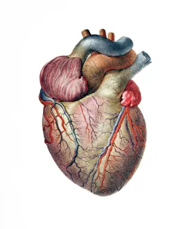
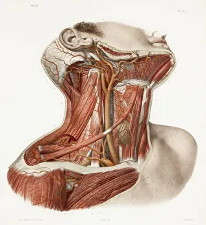
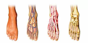
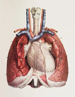
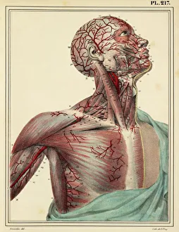
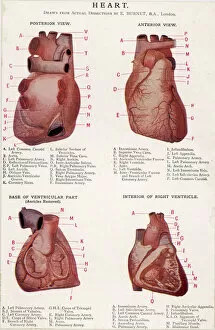
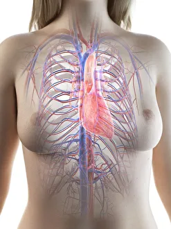
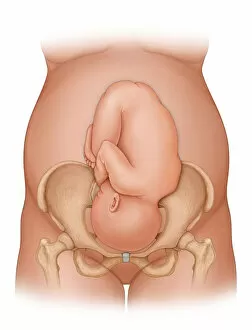
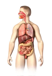
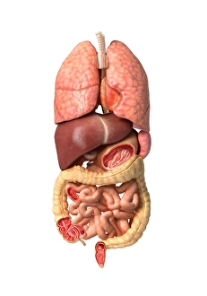
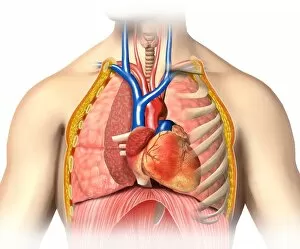
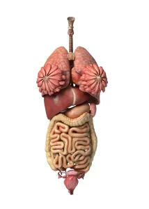
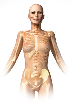
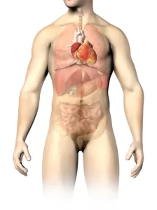
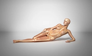
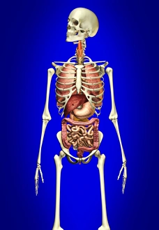
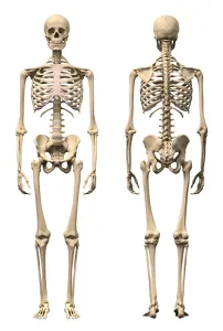
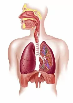
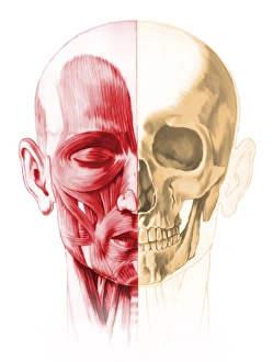
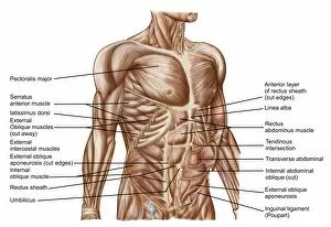
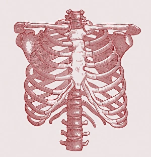
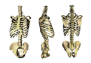
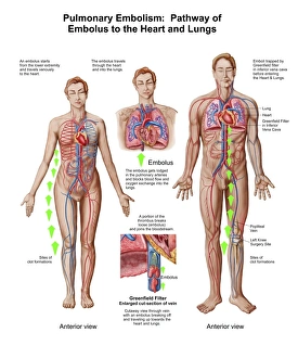
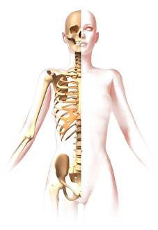
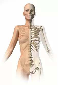
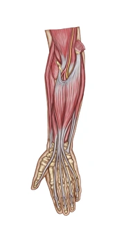
Exploring the intricate beauty of the anterior: From the delicate heart to neck vascular anatomy, historical artwork unveils its secrets. X-ray reveals the complex network of arteries in the neck and shoulder, while a detailed human foot anatomy showcases skin, veins, arteries, muscles, and bones. The interplay between heart and lungs is captured in stunning visuals. Journey back to 1825 with head and chest artery artwork. Marvel at four views of the magnificent heart itself. Dive into the molecular world with a glimpse of human growth hormone molecule. Discover female heart anatomy through captivating illustrations. Witness the miracle of life as a front view of a nine-month pregnant woman prepares for delivery with her baby phantomed within. Uncover male respiratory system and internal organs' intricacies through anatomical exploration. Finally, explore female body anatomy alongside its internal wonders - an awe-inspiring journey into our anterior realm.