
Carpals Collection
"Carpals: Unveiling the Intricacies of Hand Anatomy Across Mammals" In a mesmerizing journey through time and species


All Professionally Made to Order for Quick Shipping
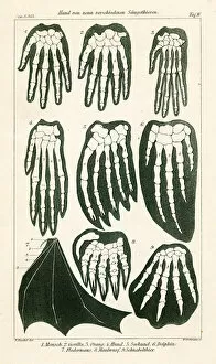
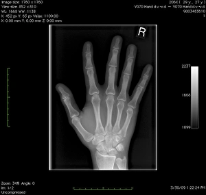
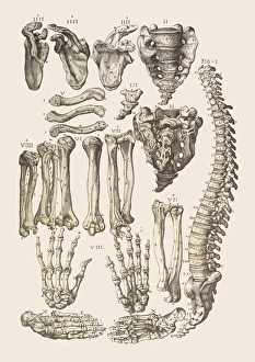
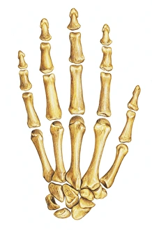
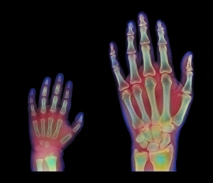
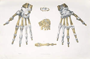
"Carpals: Unveiling the Intricacies of Hand Anatomy Across Mammals" In a mesmerizing journey through time and species, we delve into the captivating world - the small yet mighty bones that shape our hands. Comparing these remarkable structures in nine diverse mammals, a vivid 1898 color lithograph unveils their fascinating variations. From a normal hand to an intricate digital X-ray, we witness the hidden intricacies within our own human skeleton. An illustration of right hand bones guides us through each delicate component, shedding light on their individual roles in forming our dexterous grip. Moving beyond mere bone structure, wrist joint anatomy comes alive with breathtaking artwork. The interplay between ligaments and tendons is beautifully depicted, showcasing how they work harmoniously to provide stability and flexibility to this vital joint. Venturing further up the arm, detailed artwork reveals the complexity of forearm muscles. Every sinewy fiber contributes to our ability to manipulate objects with precision and strength – a testament to nature's exquisite design. However, not all tales are ones of perfection; wrist pain takes center stage in another compelling artwork. Through C013 / 8822's brushstrokes, we gain insight into common afflictions that can plague this crucial area – reminding us of its vulnerability amidst its incredible functionality. But let us not forget those who came before us - Homo neanderthalensis or Neanderthal Man. Skeleton models transport us back in time as we explore their carpals' similarities and differences from ours. These ancient beings left behind remnants that continue to intrigue scientists today. As we conclude this captivating exploration into carpals' wonders across species and eras alike, one thing becomes abundantly clear: these seemingly insignificant bones hold immense significance in shaping both our physical abilities and evolutionary history.