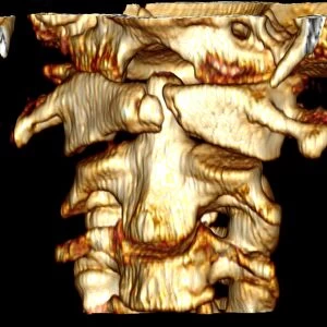
Fractured atlas vertebra, 3D CT scan

![]()
Wall Art and Photo Gifts from Science Photo Library
Fractured atlas vertebra, 3D CT scan
Fractured atlas vertebra. Coloured 3D CT (computed tomography) scan of a fractured atlas vertebra (break at upper centre) of a 34 year old man. Front is at left and part of the skull can be seen at top. The atlas vertebra (or C1) is the topmost vertebra (spinal bone) that, along with the C2 vertebra, forms the joint connecting the skull and the spine. For a picture of a second fracture in the atlas vertebra of the same patient see M330/1707
Science Photo Library features Science and Medical images including photos and illustrations
Media ID 6424041
© DU CANE MEDICAL IMAGING LTD/SCIENCE PHOTO LIBRARY
Atlas Break Broken Cervical Computed Tomography Ct Scan Fracture Injured Injury Joint Neck Osteology Profile Scanner Skeletal Spinal Column Thirties Three Dimensional Vertebra Vertebral Broken Neck Condition Disorder False Coloured Health Care Topmost Vertebrae
MADE IN THE USA
Safe Shipping with 30 Day Money Back Guarantee
FREE PERSONALISATION*
We are proud to offer a range of customisation features including Personalised Captions, Color Filters and Picture Zoom Tools
SECURE PAYMENTS
We happily accept a wide range of payment options so you can pay for the things you need in the way that is most convenient for you
* Options may vary by product and licensing agreement. Zoomed Pictures can be adjusted in the Cart.