Fraud Blocker Skip to main content

Home > Popular Themes > DNA

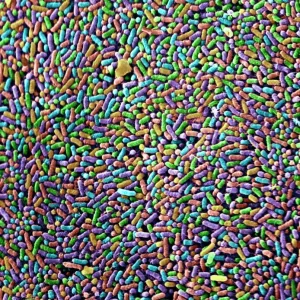
Dividing yeast cells, SEM

 : Dividing yeast cells, SEM
Sold 2
In Stock
We accept all major payment options so you can pay for the things you need in the way that is most convenient for you

Choose Your Product


Science Photo Library Photo Prints and Wall Art

Dividing yeast cells, SEM

Dividing yeast cells. Coloured scanning electron micrograph (SEM) of Schizosaccharomyces pombe yeast cells dividing. S. pombe is a single-celled fungus that is studied widely as a model organism for eukaryotic cell division. It is a rod-shaped yeast that grows by elongation at its ends. It replicates by binary fission. When it reaches a certain size its genetic material (deoxyribonucleic acid, DNA) separates to opposite ends of the cell and a division septum (wall) grows across the centre of the cell, dividing it into two daughter cells that are identical to the parent cell. Magnification: x5500 when printed 10cm wide. Science Photo Library features Science and Medical images including photos and illustrations. © STEVE GSCHMEISSNER/SCIENCE PHOTO LIBRARY

Media ID 6291947

Asexual Binary Fission Dividing Division Eukaryote Eukaryotic Eumycota Fungal Fungi Fungus Model Organism Mycology Naturemycology Re Production Replicating Replication Reproducing Single Celled Yeast Cells False Coloured Micro Biology Microbiological

FEATURES IN THESE COLLECTIONS

> Popular Themes > DNA

> Science > SEM

SECURE PAYMENTS
We accept all major payment options so you can pay for the things you need in the way that is most convenient for you

Made in USA
Safe Shipping, Easy Returns

FREE PERSONALISATION
We are proud to offer a range of customisation including Cropping, Captions, and Filters. Zoom Cropping can be adjusted in the basket


EDITORS COMMENTS

In this coloured scanning electron micrograph (SEM), we witness the intricate process of cell division in Schizosaccharomyces pombe, a single-celled fungus renowned as a widely studied model organism for eukaryotic cell division. The rod-shaped yeast, S. pombe, grows by elongation at its ends and replicates through binary fission. As the yeast cell reaches a certain size, its genetic material (DNA) begins to separate to opposite ends of the cell. The division septum, a thin wall, then grows across the centre of the cell, dividing it into two identical daughter cells. This process, known as medial fission, is a hallmark of S. pombe and is a crucial aspect of its asexual replication. This stunning SEM image, with a magnification of x5500 when printed 10cm wide, offers a mesmerizing glimpse into the microscopic world of biology. The false colours enhance the visualization of the intricate details of the dividing yeast cells, revealing the complex structures involved in the process of cell division. S. pombe is a significant organism in various fields of study, including microbiology, mycology, and eukaryote biology. Its unique mode of binary fission and the ease with which it can be cultured and manipulated make it an invaluable tool for researchers investigating the fundamental mechanisms of cell division in eukaryotes. This image serves as a testament to the beauty and complexity of nature, showcasing the intricacies of cellular processes that underpin all life on Earth.


Framed Prints of Dividing yeast cells, SEM

Discover the intricacy of life with our Media Storehouse Framed Prints featuring the captivating image of Dividing Yeast Cells. This coloured Scanning Electron Micrograph (SEM) by Science Photo Library showcases Schizosaccharomyces pombe yeast cells in the midst of division, offering a mesmerizing glimpse into the microscopic world. An excellent addition to any laboratory, classroom, or home decor, this print celebrates the beauty of science and the wonders of cellular biology.


Photo Prints of Dividing yeast cells, SEM

Discover the intricacies of nature with Media Storehouse's Photographic Prints featuring the captivating image of Dividing Yeast Cells by Science Photo Library. Witness the mesmerizing process of Schizosaccharomyces pombe yeast cells dividing in stunning detail through this Coloured Scanning Electron Micrograph (SEM). Ideal for scientific laboratories, educational institutions, or any space seeking to inspire curiosity and wonder. Bring the beauty of science into your home or office with this unique and thought-provoking addition to your decor.


Poster Prints of Dividing yeast cells, SEM

Discover the fascinating world of microbiology with our Media Storehouse Poster Prints featuring the captivating image of Dividing Yeast Cells. This coloured Scanning Electron Micrograph (SEM) by Science Photo Library showcases Schizosaccharomyces pombe yeast cells in the intricate process of division. As a single-celled fungus widely studied as a model organism, these dividing cells offer a unique glimpse into the complexities of life. Bring this stunning scientific image into your home or office to inspire curiosity and ignite discussions.


Jigsaw Puzzles of Dividing yeast cells, SEM

Discover the fascinating world of microbiology with Media Storehouse's educational Jigsaw Puzzles. This intriguing puzzle showcases Dividing Yeast Cells, captured in stunning detail through Science Photo Library's Coloured Scanning Electron Micrograph (SEM). Explore the complex process of cell division in Schizosaccharomyces pombe, a single-celled fungus widely studied as a model organism. Engage learners in a hands-on, interactive experience that fosters critical thinking and problem-solving skills while deepening their understanding of biology. Puzzle pieces are made from high-quality materials for a premium feel and easy assembly. Bring the beauty of science into your classroom or home with Media Storehouse Jigsaw Puzzles.

diamond
Made With High Grade Materials
thumb_up
Professional Quality Finish
package_2
Carefully Packed To Aid Safe Arrival

You May Also Like


USA, Washington D. C. The Washington Monument framed by cherry blossomsUSA, Washington D. C. The Washington Monument framed by cherry blossoms
Add to Basket
Sphere cover - German retreat from Lille, 1918Sphere cover - German retreat from Lille, 1918
Add to Basket
New Years Day with a Highland RegimentNew Years Day with a Highland Regiment
Add to Basket
Sphere cover - The Kaiser at Nish by Matania, WW1Sphere cover - The Kaiser at Nish by Matania, WW1
Add to Basket
Still life (hare, duck, bottles, bread and cheese), 1742 (oil on canvas)Still life (hare, duck, bottles, bread and cheese), 1742 (oil on canvas)
Add to Basket
Heather -Calluna vulgaris-, flowering, and Common Juniper -Juniperus communis-, Totengrund Valley, Wilsede, Lueneburg Heath nature reserve, Lower Saxony, GermanyHeather -Calluna vulgaris-, flowering, and Common Juniper -Juniperus communis-, Totengrund Valley, Wilsede, Lueneburg Heath nature reserve, Lower Saxony, Germany
Add to Basket
East end of Notre-Dame Cathedral, Bayeux, Normandy, France, EuropeEast end of Notre-Dame Cathedral, Bayeux, Normandy, France, Europe
Add to Basket
Rangers 2-1 Celtic: Glorious Victory (March 16, 2003)Rangers 2-1 Celtic: Glorious Victory (March 16, 2003)
Add to Basket
Chives leaf stoma, SEM C016 / 8060Chives leaf stoma, SEM C016 / 8060
Add to Basket
Chelsea Soccer Team at Stamford Bridge - Bobby SmithChelsea Soccer Team at Stamford Bridge - Bobby Smith
Add to Basket
Citrus fruitsCitrus fruits
Add to Basket
Salmonella bacteria, SEMSalmonella bacteria, SEM
Add to Basket

Gallery Wall Inspiration

Watson and Crick with their DNA model
Rosalind Franklin
DNA molecule
DNA transcription, molecular model
Double-stranded RNA molecule
DNA molecule
Watson and Crick, DNA discovers
DNA molecule, artwork
The Tarbet Hotel, Loch Lomond, Caledonian Railway poster, 1920
Peebles for Health, Rest and Recreation, railway poster, 1900-1922
Computer screen showing a human genetic sequence
X and Y chromosomes
DNA molecule
Mitosis, light micrograph
Rough endoplasmic reticulum, TEM
Cell structure
Herpes virus replicating
Fingerprint scanner
Computer artwork of a beta DNA segment and spheres
Nucleotide base matrix
DNA molecule, computer model
Nucleosome molecule
DNA molecule, abstract image
DNA nucleosome, molecular model
Dividing yeast cells, SEM
E. coli bacterium


© Copyright Media Storehouse, All Rights Reserved 2003 - 2025
Facebook     Pinterest     100% Payment Secure     Reviews IO