Cell division
![]()

Wall Art and Photo Gifts from Science Photo Library
Cell division
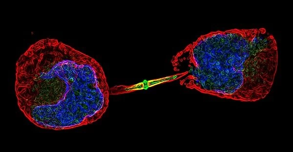
Cell division. Fluorescent micrograph of an animal cell during cytokinesis (cell division). Cytokinesis occurs after nuclear division (mitosis), which produces two daughter nuclei containing deoxyribonucleic acid (DNA, blue). A contractile ring made of actin filaments forms beneath the cells plasma membrane. This contracts, creating a furrow in the cytoplasm. The furrow deepens, separating the cytoplasm and cell organelles, until the two new cells are formed. The red structures are microtubules, part of the cells cytoskeleton, along with the actin filaments. They separate the chromosomes during mitosis
Science Photo Library features Science and Medical images including photos and illustrations
Media ID 6453797
© DR PAUL ANDREWS, UNIVERSITY OF DUNDEE/ SCIENCE PHOTO LIBRARY
Actin Filament Cell Division Contractile Ring Cytokinesis Cytological Cytology Cytoplasmic Cleavage Cytoskeletal Cytoskeleton Dividing Division Filaments Fluorescence Micrograph Furrow Hela Cells Microscope Microtubule Microtubules Mitotic Nucleus Panoramic Plasma Membrane Separating Spindle Spindles Cells Deoxyribonucleic Acid Genetics
EDITORS COMMENTS
This print captures the intricate process of cell division, showcasing a fluorescent micrograph of an animal cell during cytokinesis. The image showcases the remarkable phenomenon that occurs after nuclear division, known as mitosis, where two daughter nuclei containing DNA are produced. In this mesmerizing display, a contractile ring composed of actin filaments can be seen forming beneath the cell's plasma membrane. As it contracts, a furrow is created in the cytoplasm, gradually deepening and separating both the cytoplasm and cell organelles until two new cells are formed. The vibrant blue hue represents deoxyribonucleic acid (DNA), which plays a vital role in genetic information transfer within cells. Additionally, red structures called microtubules are visible throughout the image; they form part of the cell's cytoskeleton along with actin filaments and aid in chromosome separation during mitosis. This panoramic photograph not only highlights the biological marvels occurring at a microscopic level but also serves as a testament to our ever-expanding knowledge of cellular biology. It offers us an awe-inspiring glimpse into nature's complexity and reminds us of how essential these processes are for life itself.
MADE IN THE USA
Safe Shipping with 30 Day Money Back Guarantee
FREE PERSONALISATION*
We are proud to offer a range of customisation features including Personalised Captions, Color Filters and Picture Zoom Tools
SECURE PAYMENTS
We happily accept a wide range of payment options so you can pay for the things you need in the way that is most convenient for you
* Options may vary by product and licensing agreement. Zoomed Pictures can be adjusted in the Cart.

