
Deoxyribonucleic Acid Collection
"Unlocking the Blueprint of Life


All Professionally Made to Order for Quick Shipping
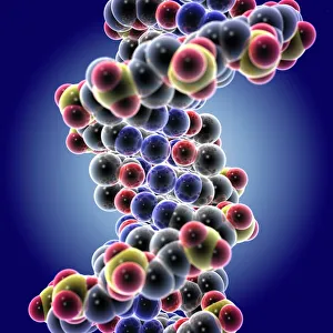
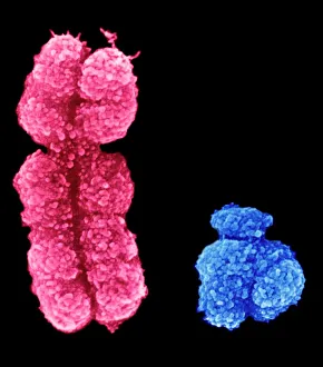
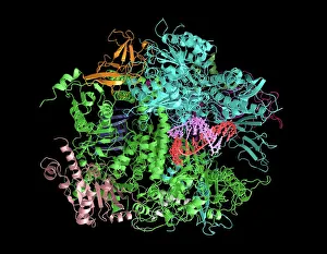
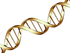
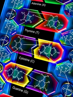
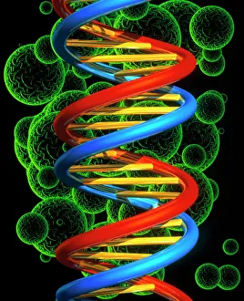
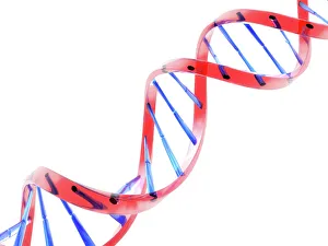
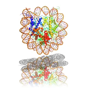
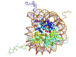
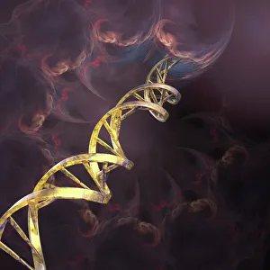
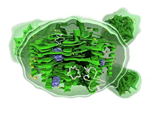
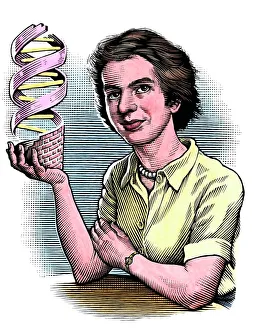
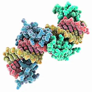
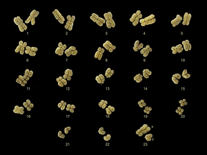
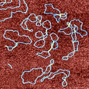
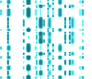
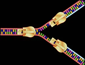
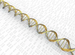
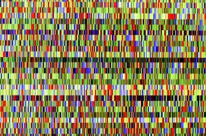
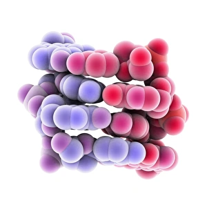
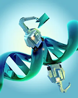
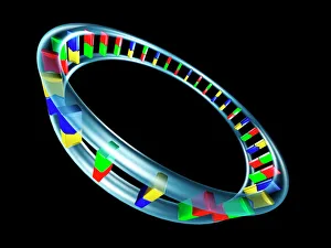
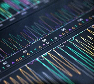
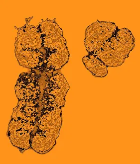
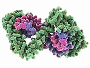
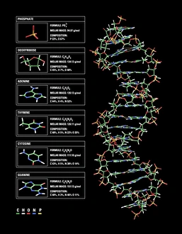
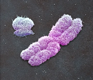
"Unlocking the Blueprint of Life: Exploring the Marvels of Deoxyribonucleic Acid" From its iconic double helix structure to its role as the carrier of genetic information, the DNA molecule stands at the core of life's intricate tapestry. The X and Y chromosomes, known for determining our biological sex, are just a fraction of what this remarkable molecule encompasses. DNA transcription, a process where genetic instructions are converted into RNA molecules, allows for protein synthesis and ultimately shapes our traits. This molecular model unveils the intricacies involved in this vital mechanism. Watson and Crick's groundbreaking discovery in 1953 revolutionized biology forever. Their unraveling of DNA's structure paved the way for countless scientific advancements that continue to shape our understanding today. Mitosis, captured through a light micrograph, showcases how DNA faithfully replicates itself during cell division. This fundamental process ensures growth and repair within living organisms. In this computer artwork featuring a beta DNA segment and spheres representing nucleotides, we witness science merging with artistry to depict both complexity and beauty entwined within every strand. The nucleotide base matrix acts as an essential foundation upon which our genetic code is built. Its precise arrangement determines everything from eye color to disease susceptibility—a testament to nature's meticulous design. A computer model showcasing the three-dimensional structure of a DNA molecule further emphasizes its elegance. Every twist and turn holds invaluable information that defines who we are at our very core. Nucleosomes—molecular complexes formed by wrapping DNA around proteins—play a crucial role in organizing chromatin structures within cells. This detailed molecular model highlights their significance in gene regulation and genome stability. An abstract image captures the essence of a DNA molecule—an enigmatic dance between orderliness and randomness that underpins all life forms on Earth. It symbolizes both unity among species yet individuality within each organism. As we delve deeper into understanding deoxyribonucleic acid, we unravel the secrets of our existence.