Fraud Blocker Skip to main content

Mitotic Collection

"Captivating Moments of Mitotic Cell Division Unveiled

Background imageMitotic Collection: Mitosis, light micrograph

Mitosis, light micrograph
Mitosis. Confocal light micrograph of the stages of mitosis (nuclear division) and cytokinesis (cell division). During mitosis the nuclear envelope disintegrates (3rd image) and the chromosomes (blue)

Background imageMitotic Collection: Cell division, fluorescent micrograph

Cell division, fluorescent micrograph
Cell division. Immunofluorescent light micrograph of a human epithelial cell (centre) during the late anaphase stage of mitosis

Background imageMitotic Collection: Dividing cells

Dividing cells. Computer artwork of a spiral of cells during mitosis (nuclear division). During mitosis two daughter nuclei are formed from one parent nucleus

Background imageMitotic Collection: Cell division, fluorescent micrograph

Cell division, fluorescent micrograph
Cell division. Immunofluorescent light micrograph of a human epithelial cell (centre) during the interphase stage of mitosis

Background imageMitotic Collection: Plant cell mitosis, light micrograph

Plant cell mitosis, light micrograph
Plant cell mitosis. Light micrograph of root tip cells from an onion (Allium sp.) undergoing cell division (mitosis). From top left to bottom right

Background imageMitotic Collection: Roundworm germ cells, light micrograph C016 / 9538

Roundworm germ cells, light micrograph C016 / 9538
Roundworm germ cells. Light micrograph of germ cells from a roundworm (Ascaris sp.), undergoing mitosis (nuclear division)

Background imageMitotic Collection: Dividing cell, TEM

Dividing cell, TEM
Dividing cell. Transmission electron micrograph (TEM) of a section through a cell dividing through the process of mitosis

Background imageMitotic Collection: Dividing cancer cell, SEM C014 / 0362

Dividing cancer cell, SEM C014 / 0362
Dividing cancer cell. Coloured scanning electron micrograph (SEM) of a colorectal cancer cell undergoing mitosis (nuclear division) and splitting into two daughter cells (left and right)

Background imageMitotic Collection: Mouth cancer cell dividing, SEM

Mouth cancer cell dividing, SEM
Mouth cancer dividing. Coloured scanning electron micrograph (SEM) of a squamous cell carcinoma (cancer) cell from a human mouth undergoing mitosis (nuclear division)

Background imageMitotic Collection: Dividing liver cancer cell, SEM

Dividing liver cancer cell, SEM
Dividing liver cancer cell. Coloured scanning electron micrograph (SEM) of a hepatocellular carcinoma (HCC) cell undergoing mitosis (nuclear division)

Background imageMitotic Collection: Dividing cancer cell, SEM C014 / 0361

Dividing cancer cell, SEM C014 / 0361
Dividing cancer cell. Coloured scanning electron micrograph (SEM) of a colorectal cancer cell undergoing mitosis (nuclear division) and splitting into two daughter cells (left and right)

Background imageMitotic Collection: Dividing bone cancer cell, SEM

Dividing bone cancer cell, SEM
SEM Dividing Osteosarcoma cells Osteosarcoma is an aggressive malignant neoplasm arising from primitive transformed cells of mesenchymal origin (and thus a sarcoma)

Background imageMitotic Collection: Lung cancer cell division, artwork C013 / 4630

Lung cancer cell division, artwork C013 / 4630
Lung cancer cell division. Computer artwork of a lung cancer cell during cell division (cytokinesis). The two daughter cells remain temporarily joined by a cytoplasmic bridge (centre)

Background imageMitotic Collection: 4-cell embryo embryo, artwork

4-cell embryo embryo, artwork
4-cell embryo. Image 3 of 4. Computer model representing 4 daughter cells. The development of an embryo is called embryogenesis

Background imageMitotic Collection: Mitosis, fluorescence micrograph

Mitosis, fluorescence micrograph
Mitosis. Fluorescence micrograph of a cell (centre) in anaphase during mitosis (nuclear division). During mitosis two daughter nuclei are formed from one parent nucleus

Background imageMitotic Collection: Multi-celled embryo, artwork

Multi-celled embryo, artwork
Multi-celled embryo. Image 4 of 4. Computer model representing a cluster of daughter cells. The development of an embryo is called embryogenesis

Background imageMitotic Collection: Single-celled embryo, artwork

Single-celled embryo, artwork
Single-celled embryo. Image 1 of 4. Computer model representing a single cell. The development of an embryo is called embryogenesis

Background imageMitotic Collection: 2-cell embryo embryo, artwork

2-cell embryo embryo, artwork
2-cell embryo. Image 2 of 4. Computer model representing 2 daughter cells. The development of an embryo is called embryogenesis

Background imageMitotic Collection: Cytokinesis

Cytokinesis. Fluorescent micrograph of an animal cell during cytokinesis (cell division). Cytokinesis occurs after nuclear division (mitosis), which produces two daughter nuclei

Background imageMitotic Collection: Abnormal mitosis

Abnormal mitosis
Mitosis. Fluorescence micrograph of a cell during abnormal anaphase of mitosis (nuclear division). During mitosis two daughter nuclei are formed from one parent nucleus

Background imageMitotic Collection: Dividing cell

Dividing cell. Differential interference contrast (DIC) light micrograph of a cell (lower left) in the metaphase stage of mitosis (cell division). The cells nuclei are stained with fluorescent dye

Background imageMitotic Collection: Mitosis, TEM

Mitosis, TEM
Mitosis, coloured transmission electron micrograph (TEM). Longitudinal section through a human T cell in metaphase. During mitosis two daughter nuclei are formed from one parent nucleus

Background imageMitotic Collection: Cell mitosis

Cell mitosis. Confocal fluorescence light micrograph composite showing 6 stages of mitotic cell division. At far left, the cell has completed the first stage of cell division known as interphase in

Background imageMitotic Collection: Cell division

Cell division. Fluorescent micrograph of an animal cell during cytokinesis (cell division). Cytokinesis occurs after nuclear division (mitosis)

Background imageMitotic Collection: Dividing cell, light micrograph

Dividing cell, light micrograph
Cell division. Fluorescent light micrograph of a cell that has divided by mitosis, the asexual replication of a cell into two new cells

Background imageMitotic Collection: Mitosis

Mitosis. Fluorescence micrograph of a cell during prometaphase of mitosis (nuclear division). During mitosis two daughter nuclei are formed from one parent nucleus

Background imageMitotic Collection: Mitosis, artwork

Mitosis, artwork

Background imageMitotic Collection: DNA

DNA
Deoxyribonucleic acid. Conceptual artwork of DNA (deoxyribonucleic acid) spelt out with fluoresence micrographs of anaphase stage cells. Anaphase is a stage of mitosis (nuclear division)

Background imageMitotic Collection: Cell division, artwork

Cell division, artwork
Cell division. Computer artwork of an animal cell undergoing mitosis (nuclear division) and cytokinesis (cell division) to form two identical daughter cells (bottom)


All Professionally Made to Order for Quick Shipping

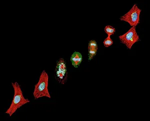
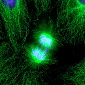
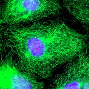
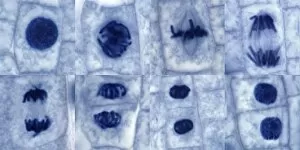
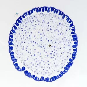
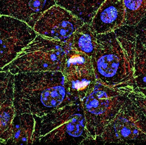
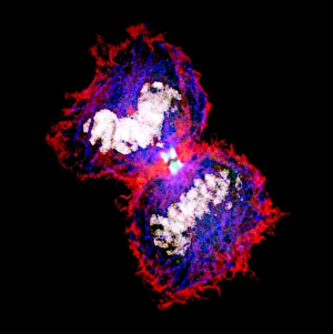
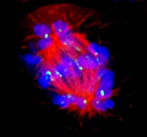
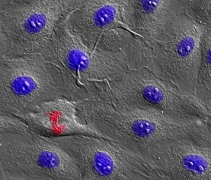
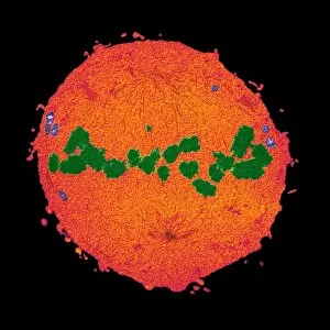
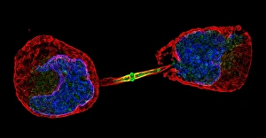
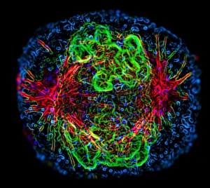
"Captivating Moments of Mitotic Cell Division Unveiled: A Glimpse into the World of Cellular Reproduction" Witness the intricate dance of life as cells divide and multiply, captured through various microscopic lenses. From light micrographs to fluorescent images, these snapshots offer a fascinating glimpse into the process known as mitosis. In one frame, observe the mesmerizing beauty of cell division under a light micrograph. The vibrant hues highlight each stage of mitosis, showcasing how cells meticulously replicate themselves to ensure growth and repair within an organism. Switching gears to fluorescent micrographs, we delve deeper into this captivating world. Fluorescent dyes illuminate dividing cells with stunning precision, revealing their inner workings like never before. Witness the remarkable symphony as chromosomes align and separate during cellular reproduction. Plant cell mitosis takes center stage in another light micrograph; here, nature's own blueprint for growth unfolds before our eyes. Delicate structures undergo precise division to create new plant tissues and enable continuous development. Even cancer cells reveal their secrets through scanning electron microscopy (SEM). Dividing liver cancer cells are magnified in astonishing detail – their abnormal proliferation becoming evident under close examination. These striking SEM images shed light on the complex nature of cancer progression. Roundworm germ cells also make an appearance in this visual journey. Through yet another set of light micrographs, witness these tiny organisms undergoing mitotic division with astounding precision and efficiency – a testament to life's resilience across species. Zooming even further down at a cellular level using transmission electron microscopy (TEM), we uncover intricate details that were once hidden from view. Dividing cells come alive in high resolution; organelles rearrange themselves while DNA replicates faithfully within each nucleus. Lastly, explore mouth cancer cell division through SEM imagery - a stark reminder that even our own bodies can succumb to uncontrolled replication processes gone awry. These powerful visuals serve as both cautionary tales and inspiration for ongoing research in the fight against cancer.