Home > Popular Themes > Human Body
Cell mitosis
![]()

Wall Art and Photo Gifts from Science Photo Library
Cell mitosis
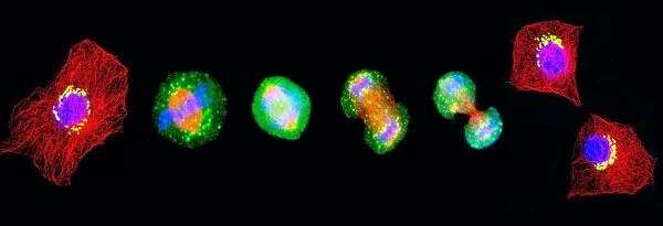
Cell mitosis. Confocal fluorescence light micrograph composite showing 6 stages of mitotic cell division. At far left, the cell has completed the first stage of cell division known as interphase in which the chromosome number is doubled. During, prophase, spindle fibres (red) form as the nuclear membrane breaks down. At metaphase (second from left), the chromosomes (blue) align at the equator of the cell as the spindles pull the two sets of chromosomes apart during anaphase (third from left). During telophase, the cell begins to pinch in and divide (fifth from left) to break apart into two daughter cells (far right). The golgi apparatus is stained green. Magnification: x300 when printed 10cm wide
Science Photo Library features Science and Medical images including photos and illustrations
Media ID 6453827
© THOMAS DEERINCK, NCMIR/SCIENCE PHOTO LIBRARY
Anaphase Cell Division Cell Nucleus Chromosome Chromosomes Daughter Cells Dividing Golgi Apparatus Golgi Body Interphase Metaphase Microtubule Microtubules Mitosis Mitotic Prophase Telophase Deoxyribonucleic Acid Light Micrograph Micro Biology Microbiological
EDITORS COMMENTS
This print captures the intricate process of cell mitosis, showcasing the six stages of mitotic cell division in stunning detail. At first glance, one can observe the remarkable transformation that occurs within a single cell as it undergoes this essential biological process. Starting from the far left, we witness interphase, where the chromosome number is doubled and sets the stage for division. Moving along, prophase reveals an awe-inspiring sight as spindle fibers (depicted in red) form while the nuclear membrane breaks down. Metaphase follows suit with chromosomes aligning at the equator of the cell, preparing for their separation during anaphase (third from left). The dynamic nature of this phase is evident as spindles pull apart two sets of chromosomes. As telophase ensues (fifth from left), a sense of anticipation fills the air. The cell begins to pinch inwards and divide itself into two distinct daughter cells (far right), each carrying its own set of genetic information. Throughout this mesmerizing journey through cellular life cycles, green-stained golgi apparatus stands out amidst all other structures. With a magnification level of x300 when printed 10cm wide, this micrograph composite offers viewers an up-close look at these microscopic wonders that shape our existence. It serves as a testament to both scientific curiosity and artistic appreciation for nature's hidden beauty within our very own bodies.
MADE IN THE USA
Safe Shipping with 30 Day Money Back Guarantee
FREE PERSONALISATION*
We are proud to offer a range of customisation features including Personalised Captions, Color Filters and Picture Zoom Tools
SECURE PAYMENTS
We happily accept a wide range of payment options so you can pay for the things you need in the way that is most convenient for you
* Options may vary by product and licensing agreement. Zoomed Pictures can be adjusted in the Cart.


