
Premium Framed Print : Animal cell anatomy, diagram


![]()
Framed Photos From Science Photo Library
Animal cell anatomy, diagram
Animal cell anatomy. Diagram showing the internal and external anatomy of an animal cell
Science Photo Library features Science and Medical images including photos and illustrations
Media ID 6317819
© FRANCIS LEROY, BIOCOSMOS/SCIENCE PHOTO LIBRARY
Animal Cell Cell Biology Cell Membrane Cellular Centrosome Chromatin Cut Away Cytosol Diagram Endoplasmic Reticulum Internal Lysosome Mitochondrion Nuclear Membrane Nuclear Pore Nucleolus Nucleus Organelle Organelles Plasma Membrane Ribosome Ribosomes Smooth Endoplasmic Reticulum Exocytosis
14"x16" Premium Frame
Contemporary style Premium Wooden Frame with 8"x10" Print. Complete with 2" White Mat and 1.25" thick MDF frame. Printed on 260 gsm premium paper. Glazed with shatter proof UV coated acrylic glass. Backing is paper covered backing with rubber bumpers. Supplied ready to hang with a pre-installed sawtooth/wire hanger. Care Instructions: Spot clean with a damp cloth. Securely packaged in a clear plastic bag and envelope in a reinforced cardboard shipper
FSC Real Wood Frame and Double Mounted with White Conservation Mountboard - Professionally Made and Ready to Hang
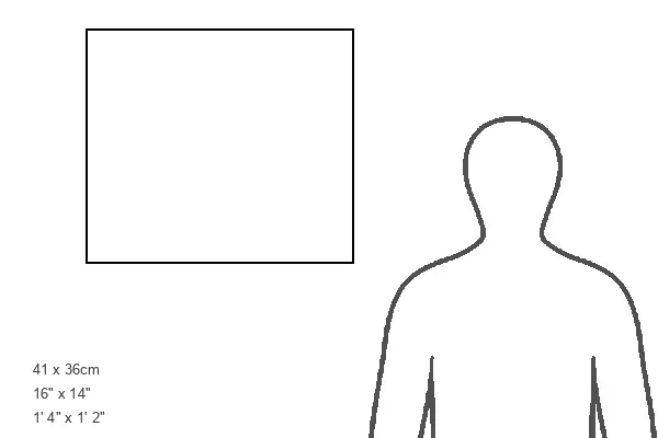
Estimated Image Size (if not cropped) is 25.4cm x 20.3cm (10" x 8")
Estimated Product Size is 40.6cm x 35.6cm (16" x 14")
These are individually made so all sizes are approximate
Artwork printed orientated as per the preview above, with landscape (horizontal) orientation to match the source image.
EDITORS COMMENTS
This print from Science Photo Library showcases the intricate anatomy of an animal cell. With meticulous detail, the diagram presents both the internal and external features of this essential building block of life. The artwork beautifully captures the complexity and elegance found within a single cut-away view. The image allows us to explore the fascinating world inside an animal cell, revealing its various organelles and structures that play crucial roles in cellular function. From the nucleus, which houses genetic material and controls cellular activities, to mitochondria responsible for energy production, each component is depicted with precision. Highlighted in vibrant colors against a dark background, we can observe key elements such as ribosomes involved in protein synthesis, endoplasmic reticulum facilitating transportation within cells, lysosomes responsible for waste disposal, and plasma membrane acting as a protective barrier. This illustration serves as a visual feast for biology enthusiasts or anyone seeking to understand the intricacies of life at a microscopic level. It exemplifies how art can merge seamlessly with science to create educational masterpieces that inspire curiosity and appreciation for our biological existence.
MADE IN THE USA
Safe Shipping with 30 Day Money Back Guarantee
FREE PERSONALISATION*
We are proud to offer a range of customisation features including Personalised Captions, Color Filters and Picture Zoom Tools
SECURE PAYMENTS
We happily accept a wide range of payment options so you can pay for the things you need in the way that is most convenient for you
* Options may vary by product and licensing agreement. Zoomed Pictures can be adjusted in the Basket.


![(15) [Gate of all Nations, Persepolis, Fars], 1840s-60s. Creator: Luigi Pesce](/sq/731/15-gate-all-nations-persepolis-fars-20171901.jpg.webp)