Pillow > Science Photo Library > Specialist Imaging
Pillow : Thale cress stigma, micrograph


![]()
Home Decor from Science Photo Library
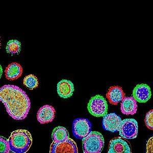
Thale cress stigma, micrograph
Thale cress stigma. Fluorescence micrograph of a stigma (female reproductive structure) from a thale cress (Arabidopsis thaliana) flower. A fluorescence microscope is an optical microscope that uses fluorescence and phosphorescence instead of, or in addition to, reflection and absorption - as used by normal light microscopes
Science Photo Library features Science and Medical images including photos and illustrations
Media ID 6328599
© HEITI PAVES/SCIENCE PHOTO LIBRARY
Arabidopsis Thaliana Carpel Colourful Fluorescence Micrograph Fluorescence Microscope Fluorescent Microscopy Mouse Ear Cress Organs Parts Reproductive Part Reproductive Structure Stigma Structures Thale Cress Gynoecium Light Micrograph Light Microscope
18"x18" (46x46cm) Pillow
18"x18" (46x46cm) Faux Suede Pillow with a plush soft feel. Your choice of image fills the front, with a stone colored faux suede back. Flat sewn concealed white zip.
Accessorise your space with decorative, soft pillows
Estimated Product Size is 45.7cm x 45.7cm (18" x 18")
These are individually made so all sizes are approximate
Artwork printed orientated as per the preview above, with landscape (horizontal) or portrait (vertical) orientation to match the source image.
EDITORS COMMENTS
This print showcases the intricate beauty of a thale cress stigma, captured through a fluorescence microscope. Set against a striking black background, the image reveals an explosion of vibrant colors that highlight the delicate features of this female reproductive structure. The thale cress, scientifically known as Arabidopsis thaliana, is a small flowering plant widely used in botanical research due to its genetic simplicity and fast growth cycle. The microscopic view offers an unprecedented glimpse into the world of botany and biology, unveiling the anatomical details of this essential organ within angiosperms. The fluorescent lighting technique employed in this micrograph enhances our understanding by illuminating specific structures and organs not visible under normal light microscopy. As we delve into this mesmerizing composition, we are reminded of nature's remarkable complexity and diversity. Each component plays a crucial role in plant reproduction, with the stigma acting as one part of the gynoecium or female reproductive system. This image serves as both an artistic masterpiece and scientific marvel - bridging aesthetics with knowledge. Science Photo Library presents us with yet another extraordinary piece that invites contemplation on life's intricacies while celebrating the wonders found within flora. It serves as a reminder that even at microscopic levels, there is immense beauty waiting to be explored and understood.
MADE IN THE USA
Safe Shipping with 30 Day Money Back Guarantee
FREE PERSONALISATION*
We are proud to offer a range of customisation features including Personalised Captions, Color Filters and Picture Zoom Tools
SECURE PAYMENTS
We happily accept a wide range of payment options so you can pay for the things you need in the way that is most convenient for you
* Options may vary by product and licensing agreement. Zoomed Pictures can be adjusted in the Cart.