Pillow : Aortic aneurysm


![]()
Home Decor From Science Photo Library
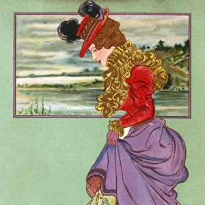
Aortic aneurysm
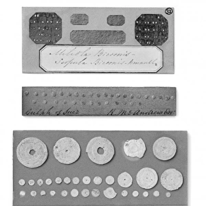
Aortic aneurysm. Artwork from Jean Cruveilhiers medical textbook Anatomie pathalogique du corps humain, published 1835. The two large illustrations are views of an aortic aneurysm (brown, centre), or bulging of the aorta caused by a weakening of its outer wall. Local blood pressure inflates the weakened artery. The heart has been dissected away to reveal the aneurysm. Aneurysms can burst at any time, causing instant death, and the larger the bulge the greater the danger. The third, smaller illustration on the right is a cross-section of myocardium, or heart muscle. Cruveilhier (1791- 1874) was an eminent French anatomist
Science Photo Library features Science and Medical images including photos and illustrations
Media ID 6419341
© MEHAU KULYK/SCIENCE PHOTO LIBRARY
1835 Aneurysm Aorta Aortic Arterial Bulge Bulging Dissected Dissection Heart Muscle Historical Image Pathological Pathology Swelling Artery Condition Disorder Health Care Humain
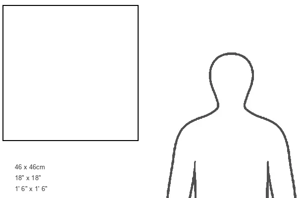
18"x18" (46x46cm) Pillow
18"x18" (46x46cm) Faux Suede Pillow with a plush soft feel. Your choice of image fills the front, with a stone colored faux suede back. Flat sewn concealed white zip.
Accessorise your space with decorative, soft pillows
Estimated Product Size is 45.7cm x 45.7cm (18" x 18")
These are individually made so all sizes are approximate
Artwork printed orientated as per the preview above, with landscape (horizontal) or portrait (vertical) orientation to match the source image.
EDITORS COMMENTS
This print showcases a historical illustration from Jean Cruveilhier's medical textbook, "Anatomie pathologique du corps humain" published in 1835. The artwork provides an intricate view of an aortic aneurysm, a condition characterized by the bulging of the aorta due to weakness in its outer wall. In this image, the heart has been meticulously dissected away to reveal the alarming presence of the aneurysm. Aneurysms pose a significant threat as they can rupture at any moment, leading to instantaneous death. The danger intensifies with larger bulges on the artery wall. This visual representation serves as both a testament to medical history and a stark reminder of the perils associated with this disorder. The smaller illustration on the right offers viewers a cross-section view of myocardium, or heart muscle tissue. Through Cruveilhier's expertise and attention to detail, we gain insight into pathological anatomy during that era. Jean Cruveilhier (1791-1874), an esteemed French anatomist, contributed immensely to our understanding of human anatomy through his groundbreaking work. His illustrations continue to be revered for their accuracy and scientific significance. This remarkable photograph from Science Photo Library not only captures our curiosity but also highlights how far medical knowledge and healthcare have progressed since these early discoveries were made in the 19th century.
MADE IN THE USA
Safe Shipping with 30 Day Money Back Guarantee
FREE PERSONALISATION*
We are proud to offer a range of customisation features including Personalised Captions, Color Filters and Picture Zoom Tools
SECURE PAYMENTS
We happily accept a wide range of payment options so you can pay for the things you need in the way that is most convenient for you
* Options may vary by product and licensing agreement. Zoomed Pictures can be adjusted in the Basket.