Fraud Blocker Skip to main content

Aneurysm Collection

Aneurysm, a medical condition that has intrigued scientists and physicians for centuries

Background imageAneurysm Collection: Rupture d un anevrysme de l aorte dans la cavite thoracique (colour litho)

Rupture d un anevrysme de l aorte dans la cavite thoracique (colour litho)
5210382 Rupture d un anevrysme de l aorte dans la cavite thoracique (colour litho) by French School, (19th century); Private Collection; (add.info)

Background imageAneurysm Collection: Jean Louis Petits machine to compress

Jean Louis Petits machine to compress the femoral artery during amputation of the thigh 1, and bandages 2, aneurysm needle 3 and scissors 4

Background imageAneurysm Collection: Model of a tumour of the left ventricle, 1799 (wax)

Model of a tumour of the left ventricle, 1799 (wax)
CHT215730 Model of a tumour of the left ventricle, 1799 (wax) by French School, (18th century); Musee Dupuytren, Paris, France; (add.info)

Background imageAneurysm Collection: Aortic aneurysm, illustration

Aortic aneurysm, illustration
Aortic aneurysm. Computer illustration showing an aneurysm in the ascending aorta

Background imageAneurysm Collection: Artist depcition of abdominal aortic aneuryism (with labels)

Artist depcition of abdominal aortic aneuryism (with labels)

Background imageAneurysm Collection: Brain aneurysm, 3D scan

Brain aneurysm, 3D scan
Brain aneurysm. 3D computed tomography (CT) angiogram coupled with a magnetic resonance imaging (MRI) scan of the brain of a 38-year-old, showing a large aneurysm (bright)

Background imageAneurysm Collection: Sir Astley Paston Cooper

Sir Astley Paston Cooper (1768 - 1841), 1st Baronet, English surgeon and anatomist, surgeon to Guys Hospital. In 1820 he removed an infected sebaceous cyst from the head of King George IV

Background imageAneurysm Collection: Abdominal aortic aneurysm, ultrasound C017 / 7649

Abdominal aortic aneurysm, ultrasound C017 / 7649
Abdominal aortic aneurysm. Doppler ultrasound scan of the abdomen of a 83 year old female patient with an abdominal aortic aneurysm (aA)

Background imageAneurysm Collection: Brain aneurysm, DSA scan

Brain aneurysm, DSA scan
Brain aneurysm. Coloured digital subtraction angiogram (DSA) of the head of a 38-year-old, showing a large aneurysm (bright, centre) of the right internal carotid artery

Background imageAneurysm Collection: Aneurysm treatment, angiogram C018 / 0537

Aneurysm treatment, angiogram C018 / 0537
Aneurysm treatment. Coloured angiogram (X-ray) overlaid on a computer model of the treatment of a 39 year old patient with an aneurysm of the posterior communicating artery

Background imageAneurysm Collection: Aneurysm treatment, angiogram C018 / 0536

Aneurysm treatment, angiogram C018 / 0536
Aneurysm treatment. Angiogram (X-ray) overlaid on a computer model of the treatment of a 39 year old patient with an aneurysm of the posterior communicating artery

Background imageAneurysm Collection: Aneurysm treatment, angiogram C018 / 0525

Aneurysm treatment, angiogram C018 / 0525
Aneurysm treatment. Coloured angiogram (X-ray) overlaid on a computer model of the treatment of a patient with an aneurysm of the internal carotid artery

Background imageAneurysm Collection: Aneurysm treatment, angiogram C018 / 0514

Aneurysm treatment, angiogram C018 / 0514
Aneurysm treatment. Coloured angiogram (X-ray) overlaid on a computer model of the treatment of a 39 year old patient with an aneurysm of the posterior communicating artery

Background imageAneurysm Collection: Aneurysm treatment, angiogram C018 / 0519

Aneurysm treatment, angiogram C018 / 0519
Aneurysm treatment. Angiogram (X-ray) overlaid on a computer model of the treatment of a patient with an aneurysm of the internal carotid artery

Background imageAneurysm Collection: Abdominal aortic aneurysm, ultrasound C017 / 7646

Abdominal aortic aneurysm, ultrasound C017 / 7646
Abdominal aortic aneurysm. Doppler ultrasound scan of the abdomen of a 83 year old female patient with an abdominal aortic aneurysm (aA)

Background imageAneurysm Collection: Pancreatitis, CT scan

Pancreatitis, CT scan
Pancreatitis. Coloured computed tomography (CT) scan of an axial section through a patients abdomen showing a false aneurysm (red) due to chronic pancreatitis. The front of the body is at top

Background imageAneurysm Collection: Aortic aneurysm

Aortic aneurysm. Artwork from Jean Cruveilhiers medical textbook Anatomie pathalogique du corps humain, published 1835. The two large illustrations are views of an aortic aneurysm (brown, centre)

Background imageAneurysm Collection: Lung aneurysm, light micrograph

Lung aneurysm, light micrograph. The lumen of the blood vessel is occluded with a large thrombus (orange). The vessel endothelium and muscle is largely replaced with dense connective tissue (red)

Background imageAneurysm Collection: Aortic artery and aneurysm, artwork

Aortic artery and aneurysm, artwork
Artwork showing the heart (upper centre) and the bodys main aortic artery (red) which forms an arch at the top of the heart before supplying blood to the lower body

Background imageAneurysm Collection: Berry aneurysm, angiogram

Berry aneurysm, angiogram
Berry aneurysm. Coloured angiogram of the brain of a patient with an aneurysm (lower centre right) in a cerebral artery. This type of aneurysm is known as a berry, or saccular, aneurysm

Background imageAneurysm Collection: Heart of King George II, engraving

Heart of King George II, engraving
The Kings heart. An engraving of the heart of King George II of Great Britain and Ireland. George II was born in 1683 and ascended the throne in 1727 on the death of his father, George I

Background imageAneurysm Collection: Stroke causes, artwork

Stroke causes, artwork
Stroke causes. Artwork showing the three causes of a stroke, a loss of brain function(s) due to an interruption in blood supply

Background imageAneurysm Collection: Abdominal aortic aneurysm, 3D CT scan

Abdominal aortic aneurysm, 3D CT scan

Background imageAneurysm Collection: Intracranial berry aneurysm, X-ray

Intracranial berry aneurysm, X-ray
Intracranial berry aneurysm, coloured X-ray. Side view of the skull of a 44-year-old patient with an intracranial berry, or saccular

Background imageAneurysm Collection: Aortic aneurysm, X-ray

Aortic aneurysm, X-ray
Aortic aneurysm. Coloured X-ray of the chest of a 68-year-old woman with an aortic aneurysm. An aneurysm is a swelling of an artery where the wall has weakened, here on the arch where the aorta

Background imageAneurysm Collection: Aortic aneurysm CT scan

Aortic aneurysm CT scan
Aortic aneurysm, coloured 3D computed tomography (CT) scan. Side view of the abdomen of a 65-year-old patient with an extended aneurysm of the aorta


All Professionally Made to Order for Quick Shipping

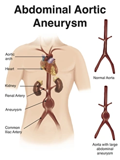
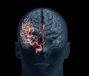
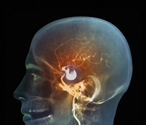
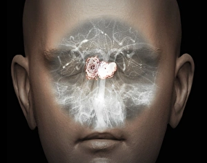
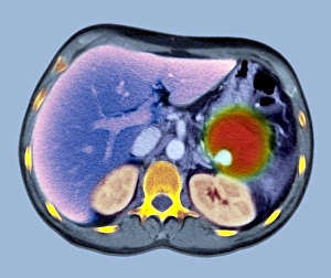
Aneurysm, a medical condition that has intrigued scientists and physicians for centuries. Sir Astley Paston Cooper, a renowned surgeon, made significant contributions to our understanding of aneurysms. His expertise in the field paved the way for advancements in treatment and diagnosis. One notable example is his work on aortic aneurysms. A striking lithograph titled "Rupture d'un anevrysme de l'aorte dans la cavite thoracique" showcases the catastrophic consequences of such an event. This visual representation serves as a reminder of the importance of early detection and intervention. Jean Louis Petit's machine to compress played a crucial role in managing aneurysms during the 18th century. This innovative device aimed to alleviate pressure within blood vessels affected by this condition, offering relief to patients suffering from its debilitating symptoms. In 1799, a wax model depicting a tumor of the left ventricle shed light on another aspect related to aneurysms. This intricate creation not only showcased anatomical details but also highlighted the potential dangers associated with these abnormal bulges in blood vessels. Moving forward, illustrations became instrumental in educating both medical professionals and individuals about different types of aneurysms. An artist's depiction vividly portrayed abdominal aortic aneurysm with labels, enabling viewers to grasp its complexities and understand potential risks involved. Advancements in technology have revolutionized our ability to diagnose and treat various forms of aneurysms effectively. Three-dimensional scans provide detailed insights into brain abnormalities caused by this condition while ultrasound imaging allows for non-invasive examination of abdominal aortic aneurysms. As we continue unraveling the mysteries surrounding aneurysms, it is essential to remember those who dedicated their lives to advancing medical knowledge like Sir Astley Paston Cooper and Jean Louis Petit. Their contributions have undoubtedly shaped modern approaches towards diagnosing, treating, and preventing this potentially life-threatening condition.