
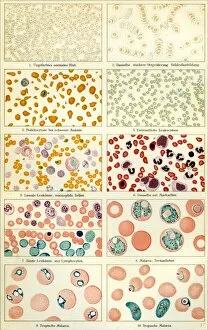
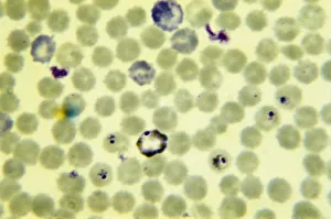
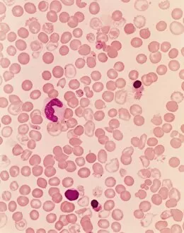
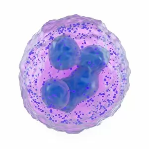
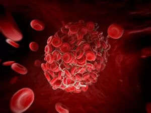
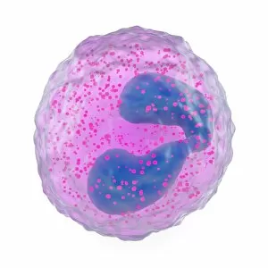
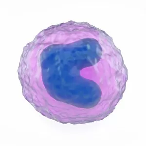
Blood Cell Collection
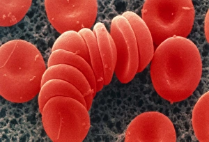
"Unveiling the Intricacies of Blood Cells: A Journey Through Microscopy" Exploring the Marvels of Stem Cells and SEM Imaging Dive into the Fascinating World Inside Veins


All Professionally Made to Order for Quick Shipping
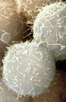
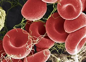
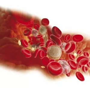
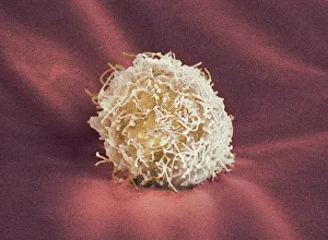
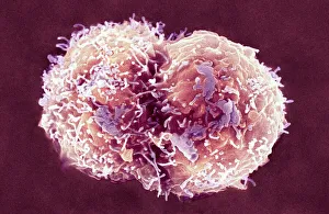
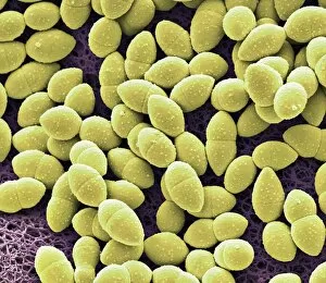
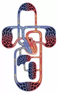
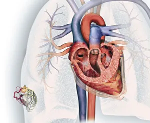
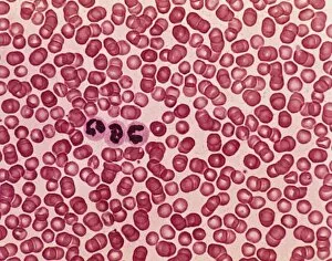
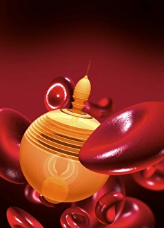
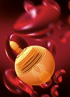
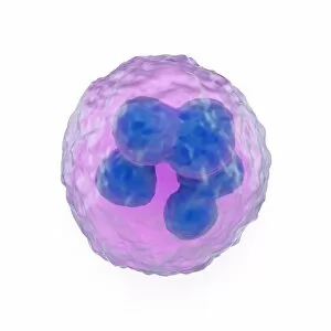
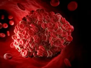
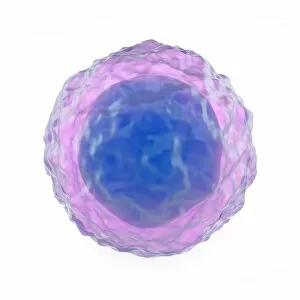
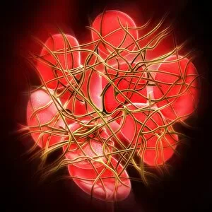
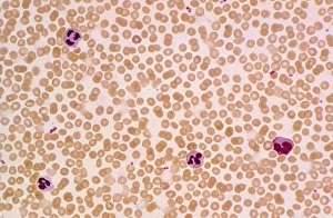
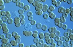
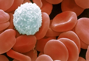
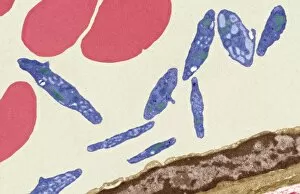
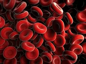
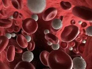
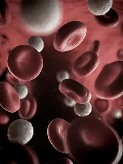
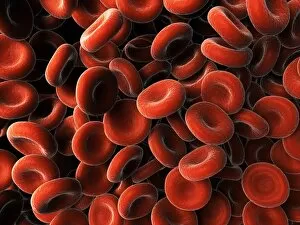
"Unveiling the Intricacies of Blood Cells: A Journey Through Microscopy" Exploring the Marvels of Stem Cells and SEM Imaging Dive into the Fascinating World Inside Veins: Diagram Unveils Bloodstream Composition Visualizing Life's Essential Components: Red, White Blood Cells, and Platelets Under SEM The Remarkable Universe of Stem Cells Revealed through Scanning Electron Microscopy (SEM) Unraveling the Mysteries Within: Witnessing White Blood Cells and Platelets in Action Medical Nanorobot Artwork Takes Us on a Mesmerizing Voyage Inside Our Bodies Peering into Nature's Hidden Gems: Discovering Diatoms and Radiolaria under SEM Captivating Close-ups of Kefir Bacteria Expose Their Unique Structures under SEM Unlocking Secrets Between Mind and Body: Diagram Explores Female Reproductive Cycle & Contraceptive Pill Effects Exploring the Intricate Beauty of Blood Cells through Scanning Electron Microscopy (SEM) Illustrating Life's Vital Elements: An Artistic Glimpse into the World of Blood Cells Journey to the Heart's Core.