Fraud Blocker Skip to main content

Blood Cell Collection (page 3)

"Unveiling the Intricacies of Blood Cells: A Journey Through Microscopy" Exploring the Marvels of Stem Cells and SEM Imaging Dive into the Fascinating World Inside Veins

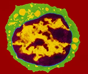
Background imageBlood Cell Collection: Coloured TEM of a T-lymphocyte white blood cell

Coloured TEM of a T-lymphocyte white blood cell

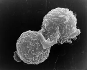
Background imageBlood Cell Collection: SEM of two polymorphonuclear white blood cells

SEM of two polymorphonuclear white blood cells
White blood cells. Scanning electron micrograph of two polymorphonuclear white blood cells (leucocytes). These cells, also known as neutrophils or polymorphs are involved in protecting the body

Background imageBlood Cell Collection: llustration of iron-deficient blood in pregnancy

llustration of iron-deficient blood in pregnancy

Background imageBlood Cell Collection: Coloured LM of blood in sickle cell anaemia

Coloured LM of blood in sickle cell anaemia
Sickle cell anaemia. Coloured light micrograph of a blood smear showing distorted red blood cells in sickle cell anaemia (drepanocytosis)

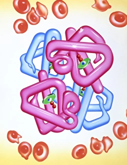
Background imageBlood Cell Collection: Iron deficient blood cells & haemoglobin molecule

Iron deficient blood cells & haemoglobin molecule
Iron-deficiency anaemia and haemoglobin. Artwork of irregularly shaped red blood cells (red) in a patient suffering from iron-deficiency anaemia

Background imageBlood Cell Collection: Medical nanorobot

Medical nanorobot. Computer illustration of a nanorobot on a T-lymphocyte (T-cell) white blood cell (green) infected by AIDS viruses (red)

Background imageBlood Cell Collection: illustration of red blood cells in thalassaemia

illustration of red blood cells in thalassaemia
Thalassaemia. Illustration of distorted red blood cells in the inherited blood disorder of thalass- aemia. Here, affected red blood cells (erythro- cytes)

Background imageBlood Cell Collection: Coloured SEM of a monocyte in a blood capillary

Coloured SEM of a monocyte in a blood capillary
Monocyte. Coloured Scanning Electron Micrograph (SEM) of a monocyte (macrophage) in a blood capillary of the " zona reticularis" of the adrenal gland

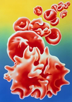
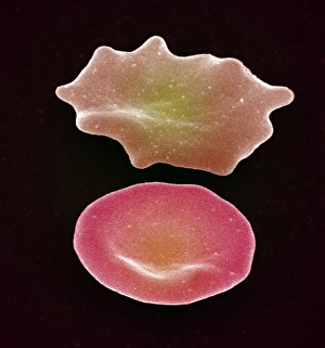
Background imageBlood Cell Collection: Red blood cell crenation, SEM

Red blood cell crenation, SEM
Red blood cell crenation. Coloured scanning electron micrograph (SEM) of two red blood cells (erythrocytes). The cell on the bottom is normal

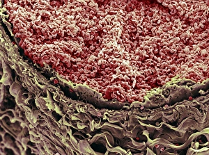
Background imageBlood Cell Collection: Red blood cells in blood vessel

Red blood cells in blood vessel
Red blood cells. Coloured scanning electron micro- graph (SEM) of red blood cells in a human blood vessel. The red blood cells (erythrocytes, at top) are filling the lumen of the blood vessel

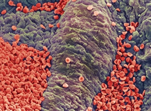
Background imageBlood Cell Collection: Coloured SEM of red blood cells on vessel wall

Coloured SEM of red blood cells on vessel wall
Red blood cells. Coloured scanning electron micrograph (SEM) of human red blood cells (erythrocytes) on the wall of a blood vessel. Red blood cells carry oxygen and carbon dioxide around the body

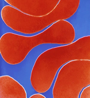
Background imageBlood Cell Collection: Colour TEM of red blood cells, rouleau formation

Colour TEM of red blood cells, rouleau formation
Red blood cells. Coloured Transmission Electron Micrograph (TEM) of sectioned human red blood cells (erythrocytes) in a rouleau formation

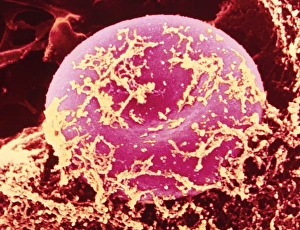
Background imageBlood Cell Collection: Coloured SEM of red blood cell covered in fibrin

Coloured SEM of red blood cell covered in fibrin
False-colour scanning electron micrograph (SEM) showing a human red blood cell (erythrocyte) with a fine covering of fibrin, the protein involved in clot formation

Background imageBlood Cell Collection: Coloured SEM of red blood cells in blood vessel

Coloured SEM of red blood cells in blood vessel
Red blood cells. Coloured scanning electron micrograph of a group of red blood cells (erythrocytes). They are travelling through a large vessel and are just entering a small vessel

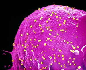
Background imageBlood Cell Collection: False-colour SEM of adenoviruses on red blood cell

False-colour SEM of adenoviruses on red blood cell
False-colour scanning electron micrograph (SEM) of a group of adenoviruses (yellow) on the surface of a red blood cell of a chicken

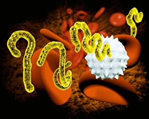
Background imageBlood Cell Collection: Ebola virus and blood cells

Ebola virus and blood cells. Computer artwork of Ebola viruses particles (yellow) attacking a human white blood cell. Red blood cells are also seen

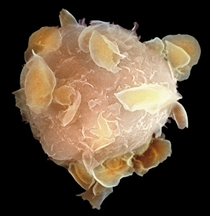
Background imageBlood Cell Collection: Bone marrow stem cell, SEM

Bone marrow stem cell, SEM
Bone marrow stem cell, coloured scanning electron micrograph (SEM). This cell is known as a multipotential stem cell because it can form the precursors to every type of blood cell

Background imageBlood Cell Collection: Blood stream, artwork

Blood stream, artwork

Background imageBlood Cell Collection: Artery, artwork

Artery, artwork

Background imageBlood Cell Collection: Arteries, artwork

Arteries, artwork

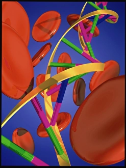
Background imageBlood Cell Collection: Computer artwork of DNA and red blood cells

Computer artwork of DNA and red blood cells
DNA & blood. Computer illustration of a molecule of DNA (Deoxyribonucleic Acid) amongst red blood cells. DNA molecules carry an organisms genetic information

Background imageBlood Cell Collection: White blood cell, artwork

White blood cell, artwork

Background imageBlood Cell Collection: Cut artery, artwork

Cut artery, artwork

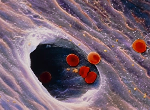
Background imageBlood Cell Collection: Ruptured venule, SEM

Ruptured venule, SEM
Ruptured venule. Coloured scanning electron micrograph (SEM) of a ruptured venule running through fatty tissue. Stacked red blood cells (rouleaux formation)

Background imageBlood Cell Collection: Blood cells, artwork

Blood cells, artwork
Blood cells. Cut-away artwork of a blood vessel showing the blood cells inside. The majority of blood cells are erythrocytes (red blood cells)

Background imageBlood Cell Collection: Red blood cells and platelets, SEM

Red blood cells and platelets, SEM
Red blood cells and platelets. Coloured scanning electon micrograph (SEM) of human erythrocytes (red blood cells) and a platelet aggregate (purple)

Background imageBlood Cell Collection: Blood cells in a kidney, SEM

Blood cells in a kidney, SEM
Blood cells in a kidney. Transmission electron micrograph (TEM) of a section through a kidney glomerulus, showing red blood cells (erythrocytes, red) and a white blood cell (leucocyte, blue)

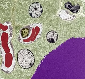
Background imageBlood Cell Collection: Thyroid follicle structures, TEM

Thyroid follicle structures, TEM
Thyroid follicle structures. Coloured transmission electron micrograph (TEM) of a section through structures in a thyroid follicle


All Professionally Made to Order for Quick Shipping

"Unveiling the Intricacies of Blood Cells: A Journey Through Microscopy" Exploring the Marvels of Stem Cells and SEM Imaging Dive into the Fascinating World Inside Veins: Diagram Unveils Bloodstream Composition Visualizing Life's Essential Components: Red, White Blood Cells, and Platelets Under SEM The Remarkable Universe of Stem Cells Revealed through Scanning Electron Microscopy (SEM) Unraveling the Mysteries Within: Witnessing White Blood Cells and Platelets in Action Medical Nanorobot Artwork Takes Us on a Mesmerizing Voyage Inside Our Bodies Peering into Nature's Hidden Gems: Discovering Diatoms and Radiolaria under SEM Captivating Close-ups of Kefir Bacteria Expose Their Unique Structures under SEM Unlocking Secrets Between Mind and Body: Diagram Explores Female Reproductive Cycle & Contraceptive Pill Effects Exploring the Intricate Beauty of Blood Cells through Scanning Electron Microscopy (SEM) Illustrating Life's Vital Elements: An Artistic Glimpse into the World of Blood Cells Journey to the Heart's Core.