Fraud Blocker Skip to main content

Blood Cell Collection (page 2)

"Unveiling the Intricacies of Blood Cells: A Journey Through Microscopy" Exploring the Marvels of Stem Cells and SEM Imaging Dive into the Fascinating World Inside Veins

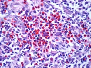
Background imageBlood Cell Collection: Hodgkins lymphoma, light micrograph

Hodgkins lymphoma, light micrograph
Hodgkins lymphoma. Light micrograph of a section through a lymph node affected by Hodgkins lymphoma, a cancer of lymphoma white blood cells. Large numbers of eosinophils (red) are present

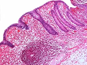
Background imageBlood Cell Collection: Appendicitis, light micrograph

Appendicitis, light micrograph
Appendicitis. Light micrograph of a section through an inflamed appendix. There is an accumulation of white blood cells (dark pink) at bottom left

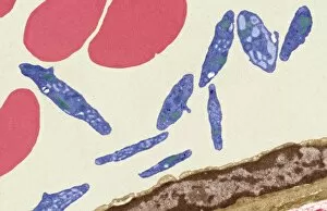
Background imageBlood Cell Collection: Blood vessel, TEM

Blood vessel, TEM
Blood vessel. Coloured transmission electron micrograph (TEM) of a section through a blood vessel, showing platelets (blue) and red blood cells (red)

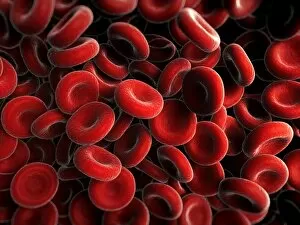
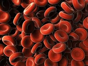
Background imageBlood Cell Collection: Red blood cells, artwork F006 / 2838

Red blood cells, artwork F006 / 2838
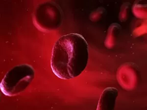
Red blood cells, computer artwork

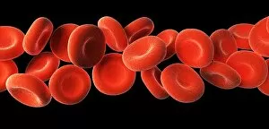
Background imageBlood Cell Collection: Red blood cells, artwork F006 / 2731

Red blood cells, artwork F006 / 2731
Red blood cells, computer artwork

Background imageBlood Cell Collection: Red blood cells, artwork F006 / 2732

Red blood cells, artwork F006 / 2732
Red blood cells, computer artwork

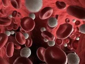
Background imageBlood Cell Collection: Blood stream, artwork F006 / 2729

Blood stream, artwork F006 / 2729
Blood stream, computer artwork

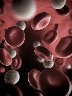
Background imageBlood Cell Collection: Blood stream, artwork F006 / 2730

Blood stream, artwork F006 / 2730
Blood stream, computer artwork

Background imageBlood Cell Collection: Red blood cells, artwork F006 / 2726

Red blood cells, artwork F006 / 2726
Red blood cells, computer artwork

Background imageBlood Cell Collection: Red blood cells, artwork F006 / 2728

Red blood cells, artwork F006 / 2728
Red blood cells, computer artwork

Background imageBlood Cell Collection: Red blood cells, artwork F006 / 2727

Red blood cells, artwork F006 / 2727
Red blood cells, computer artwork

Background imageBlood Cell Collection: Circulating insulin, artwork F006 / 2355

Circulating insulin, artwork F006 / 2355
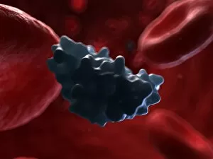
Insulin. Computer artwork of molecules of insulin circulating in the blood stream

Background imageBlood Cell Collection: Circulating insulin, artwork F006 / 2354

Circulating insulin, artwork F006 / 2354
Insulin. Computer artwork of molecules of insulin circulating in the blood stream

Background imageBlood Cell Collection: Circulating insulin, artwork F006 / 2353

Circulating insulin, artwork F006 / 2353
Insulin. Computer artwork of a molecule of insulin circulating in the blood stream

Background imageBlood Cell Collection: Red blood cells, artwork F007 / 0033

Red blood cells, artwork F007 / 0033
Red blood cells, computer artwork

Background imageBlood Cell Collection: Red blood cells, artwork F007 / 0034

Red blood cells, artwork F007 / 0034
Red blood cells, computer artwork

Background imageBlood Cell Collection: Red blood cells, artwork F007 / 0035

Red blood cells, artwork F007 / 0035
Red blood cells in a blood vessel, computer artwork

Background imageBlood Cell Collection: Red blood cells, artwork F007 / 0036

Red blood cells, artwork F007 / 0036
Red blood cells in a blood vessel, computer artwork

Background imageBlood Cell Collection: Red blood cells, artwork F006 / 8607

Red blood cells, artwork F006 / 8607
Red blood cells, computer artwork

Background imageBlood Cell Collection: Blood stream, artwork F006 / 8606

Blood stream, artwork F006 / 8606
Blood stream, computer artwork

Background imageBlood Cell Collection: Activated platelets, artwork F006 / 8056

Activated platelets, artwork F006 / 8056
Activated platelets, computer artwork

Background imageBlood Cell Collection: Activated platelets, artwork F006 / 8055

Activated platelets, artwork F006 / 8055
Activated platelets, computer artwork

Background imageBlood Cell Collection: Activated platelets, artwork F006 / 8054

Activated platelets, artwork F006 / 8054
Activated platelets, computer artwork

Background imageBlood Cell Collection: Blood stream, artwork F006 / 7964

Blood stream, artwork F006 / 7964
Blood stream, computer artwork

Background imageBlood Cell Collection: Blood stream, artwork F006 / 7963

Blood stream, artwork F006 / 7963
Blood stream, computer artwork

Background imageBlood Cell Collection: Blood stream, artwork F006 / 7962

Blood stream, artwork F006 / 7962
Blood stream, computer artwork

Background imageBlood Cell Collection: Blood stream, artwork F006 / 7961

Blood stream, artwork F006 / 7961
Blood stream, computer artwork

Background imageBlood Cell Collection: Haematopoietic stem cells, artwork

Haematopoietic stem cells, artwork
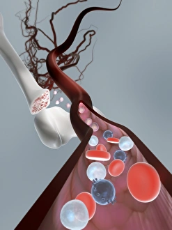
Haematopoietic stem cells. Cutaway computer artwork showing white blood cells (leucocytes, white, round), red blood cells (erythrocytes, red) and haematopoietic stem cells (HSCs)

Background imageBlood Cell Collection: Lymphocyte white blood cell, SEM C016 / 9415

Lymphocyte white blood cell, SEM C016 / 9415
Lymphocyte white blood cell. Coloured scanning electron micrograph (SEM) of a lymphocyte white blood cell. Lymphocytes are involved in the immune systems defence mechanisms, lymph system

Background imageBlood Cell Collection: Erythroblast blood cell, light micrograph

Erythroblast blood cell, light micrograph
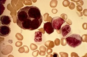
Erythroblast blood cell. Light micrograph of cells from a sample of bone marrow, including a polychromatic erythroblast (upper left). This one has multiple cell nuclei (dark red, four in total)

Background imageBlood Cell Collection: Blood cells, illustration C018 / 0802

Blood cells, illustration C018 / 0802
Blood cells. All cellular blood components originate from the same cell, the haematopoietic stem cell. The stem cell differentiates into two types of progenitor cells

Background imageBlood Cell Collection: T-lymphocyte, SEM C017 / 8306

T-lymphocyte, SEM C017 / 8306
T-Lymphocyte, coloured scanning electron micrograph (SEM). T-lymphocytes, or T-cells, are a type of white blood cell and components of the bodys immune system. They mature in the thymus

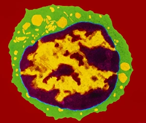
Background imageBlood Cell Collection: Colour TEM of B-lymphocyte blood cell

Colour TEM of B-lymphocyte blood cell

Background imageBlood Cell Collection: Coloured SEM of section through a human arteriole

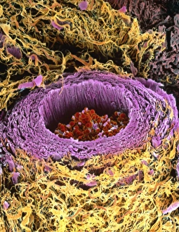
Coloured SEM of section through a human arteriole
Arteriole. Coloured scanning electron micrograph (SEM) of a cross-section through a small human artery known as an arteriole
Background imageBlood Cell Collection: Coloured TEM of a basophil white blood cell

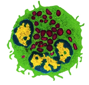
Coloured TEM of a basophil white blood cell
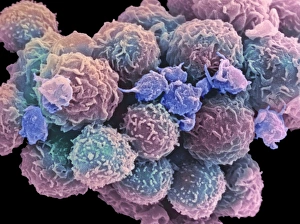
Polynuclear basophil. Coloured Transmission Electron Micrograph (TEM) of a basophil white blood cell. This basophil has one bi-lobed nucleus (yellow, lower centre) and a second nucleus at upper left
Background imageBlood Cell Collection: Electron micrograph of B-lymphocyte blood cell

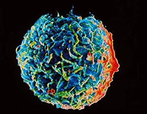
Electron micrograph of B-lymphocyte blood cell
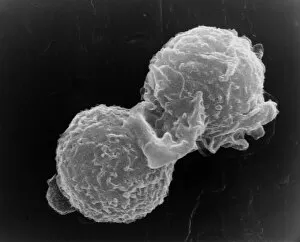
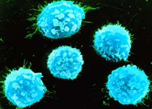
B-lymphocyte. Coloured scanning electron micrograph of a B-lymphocyte white blood cell. The cells surface is covered with tiny ridges and projections called microvilli

Background imageBlood Cell Collection: White blood cells and platelets

White blood cells and platelets
White blood cells & platelets. Coloured scanning electron micrograph (SEM) of a group of white blood cells (leucocytes) and activated platelets in human blood

Background imageBlood Cell Collection: Coloured SEM of B-lymphocyte white blood cells

Coloured SEM of B-lymphocyte white blood cells
B-lymphocyte. Coloured Scanning Electron Microgr- aph of a group of B-lymphocyte white blood cells. The cell surfaces are covered with tiny ridges and projections called microvilli

Background imageBlood Cell Collection: Platelets in a blood clot

Platelets in a blood clot. Coloured scanning electron micrograph (SEM) of platelet cells (thrombocytes) in a blood clot. They are oval shaped

Background imageBlood Cell Collection: Coloured TEM of a T-lymphocyte white blood cell

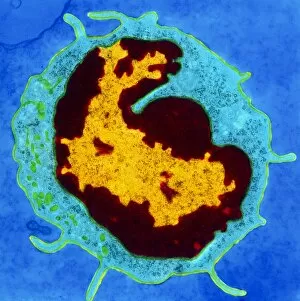
Coloured TEM of a T-lymphocyte white blood cell

Background imageBlood Cell Collection: SEM of two polymorphonuclear white blood cells

SEM of two polymorphonuclear white blood cells
White blood cells. Scanning electron micrograph of two polymorphonuclear white blood cells (leucocytes). These cells, also known as neutrophils or polymorphs are involved in protecting the body

Background imageBlood Cell Collection: llustration of iron-deficient blood in pregnancy

llustration of iron-deficient blood in pregnancy

Background imageBlood Cell Collection: Coloured LM of blood in sickle cell anaemia

Coloured LM of blood in sickle cell anaemia
Sickle cell anaemia. Coloured light micrograph of a blood smear showing distorted red blood cells in sickle cell anaemia (drepanocytosis)

Background imageBlood Cell Collection: Iron deficient blood cells & haemoglobin molecule

Iron deficient blood cells & haemoglobin molecule
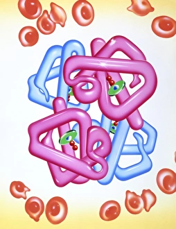
Iron-deficiency anaemia and haemoglobin. Artwork of irregularly shaped red blood cells (red) in a patient suffering from iron-deficiency anaemia

Background imageBlood Cell Collection: Medical nanorobot

Medical nanorobot. Computer illustration of a nanorobot on a T-lymphocyte (T-cell) white blood cell (green) infected by AIDS viruses (red)

Background imageBlood Cell Collection: illustration of red blood cells in thalassaemia

illustration of red blood cells in thalassaemia
Thalassaemia. Illustration of distorted red blood cells in the inherited blood disorder of thalass- aemia. Here, affected red blood cells (erythro- cytes)

Background imageBlood Cell Collection: Coloured SEM of a monocyte in a blood capillary

Coloured SEM of a monocyte in a blood capillary
Monocyte. Coloured Scanning Electron Micrograph (SEM) of a monocyte (macrophage) in a blood capillary of the " zona reticularis" of the adrenal gland

Background imageBlood Cell Collection: Red blood cell crenation, SEM

Red blood cell crenation, SEM
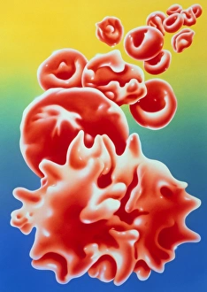
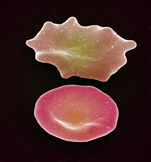
Red blood cell crenation. Coloured scanning electron micrograph (SEM) of two red blood cells (erythrocytes). The cell on the bottom is normal


All Professionally Made to Order for Quick Shipping

"Unveiling the Intricacies of Blood Cells: A Journey Through Microscopy" Exploring the Marvels of Stem Cells and SEM Imaging Dive into the Fascinating World Inside Veins: Diagram Unveils Bloodstream Composition Visualizing Life's Essential Components: Red, White Blood Cells, and Platelets Under SEM The Remarkable Universe of Stem Cells Revealed through Scanning Electron Microscopy (SEM) Unraveling the Mysteries Within: Witnessing White Blood Cells and Platelets in Action Medical Nanorobot Artwork Takes Us on a Mesmerizing Voyage Inside Our Bodies Peering into Nature's Hidden Gems: Discovering Diatoms and Radiolaria under SEM Captivating Close-ups of Kefir Bacteria Expose Their Unique Structures under SEM Unlocking Secrets Between Mind and Body: Diagram Explores Female Reproductive Cycle & Contraceptive Pill Effects Exploring the Intricate Beauty of Blood Cells through Scanning Electron Microscopy (SEM) Illustrating Life's Vital Elements: An Artistic Glimpse into the World of Blood Cells Journey to the Heart's Core.