Mouse Mat : Easter cactus stigma, SEM

![]()
Home Decor from Science Photo Library
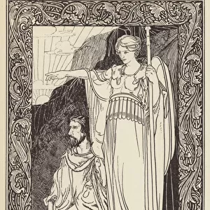
Easter cactus stigma, SEM
Easter cactus stigma. Coloured scanning electron micrograph (SEM) of part of the stigma (pink) of an Easter cactus flower (Rhipsalidopsis gaertneri). This is the top part of the female reproductive structure (carpel) of the flower. Pollen grains containing the male sex cells land on the stigma and may move down the style (not seen) into the ovary (not seen). If the male cells fertilise the female sex cells (ovules) in the ovary, then the carpel may ripen to form a fruit that contains the plants seeds. Magnification: x340 at 6x7cm size
Science Photo Library features Science and Medical images including photos and illustrations
Media ID 6288919
© SUSUMU NISHINAGA/SCIENCE PHOTO LIBRARY
Flowering Part Parts Re Production Reproductive Stigma Epiphyllum Rhipsalidopsis Gaertneri Schlumbergera
Mouse Pad
Standard Size Mouse Pad 7.75" x 9..25". High density Neoprene w linen surface. Easy to clean, stain resistant finish. Rounded corners.
Archive quality photographic print in a durable wipe clean mouse mat with non slip backing. Works with all computer mice
Estimated Product Size is 20.2cm x 23.7cm (8" x 9.3")
These are individually made so all sizes are approximate
Artwork printed orientated as per the preview above, with landscape (horizontal) or portrait (vertical) orientation to match the source image.
EDITORS COMMENTS
This print showcases the intricate beauty of an Easter cactus stigma. The coloured scanning electron micrograph (SEM) reveals a close-up view of the top part of the female reproductive structure, known as the carpel. Delicately colored in pink, this stigma serves as a landing pad for pollen grains containing male sex cells. The mesmerizing image highlights the crucial role played by the stigma in plant reproduction. As pollen lands on its surface, it may travel down the unseen style and into the ovary, which is also hidden from view. If successful fertilization occurs between male and female sex cells within the ovary's ovules, then this carpel has the potential to develop into a fruit that will bear seeds. With a magnification of x340 at 6x7cm size, this SEM photograph allows us to appreciate nature's intricate design on a microscopic scale. It provides insight into both botany enthusiasts and researchers studying plant reproduction processes. The featured flower belongs to Rhipsalidopsis gaertneri species commonly referred to as Easter cactus or Schlumbergera epiphyllum. This stunning visual representation captures not only its structural beauty but also emphasizes its importance in sustaining future generations through seed production. Science Photo Library presents this remarkable image that combines scientific exploration with artistic appreciation for nature's wonders.
MADE IN THE USA
Safe Shipping with 30 Day Money Back Guarantee
FREE PERSONALISATION*
We are proud to offer a range of customisation features including Personalised Captions, Color Filters and Picture Zoom Tools
SECURE PAYMENTS
We happily accept a wide range of payment options so you can pay for the things you need in the way that is most convenient for you
* Options may vary by product and licensing agreement. Zoomed Pictures can be adjusted in the Cart.