
Mycology Collection
Mycology: Unveiling the Hidden World of Fungi Delving into the fascinating realm of mycology, we encounter a diverse array of fungal wonders


All Professionally Made to Order for Quick Shipping
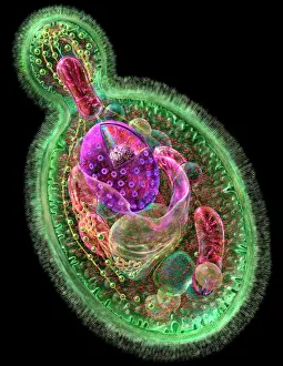
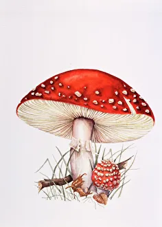
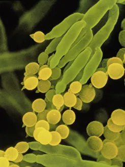
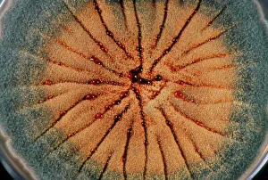
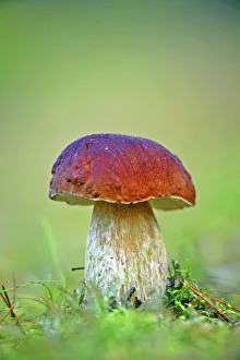
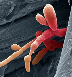
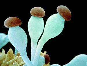
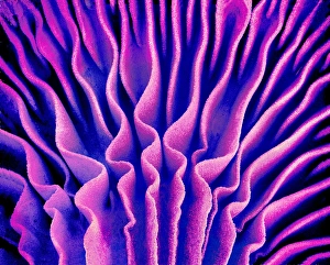
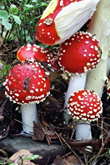
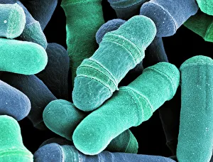
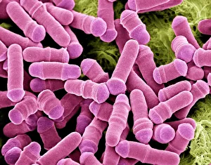
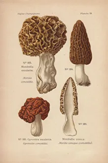
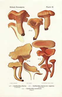
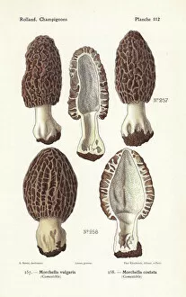
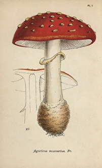
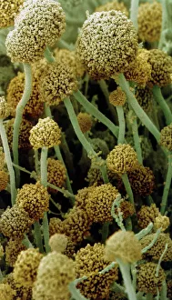
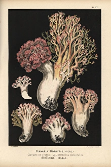
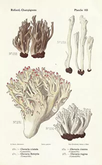
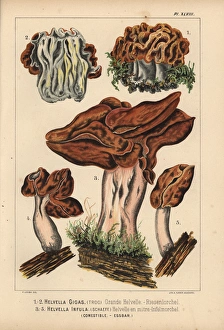
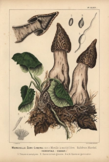
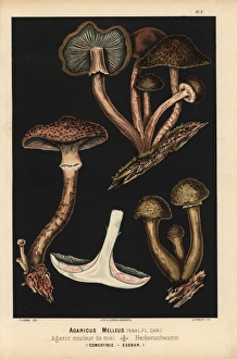
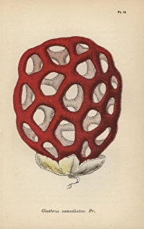
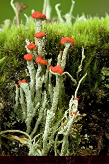
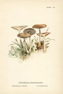
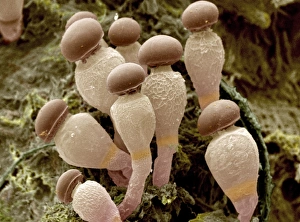
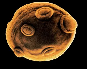
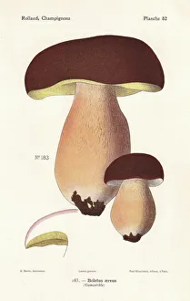
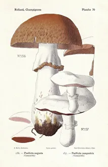
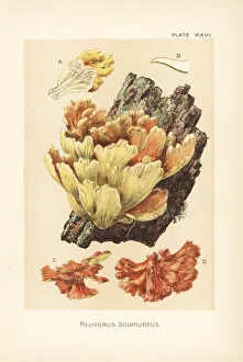
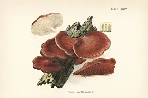
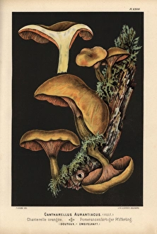
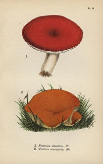
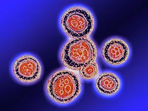
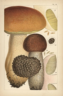
Mycology: Unveiling the Hidden World of Fungi Delving into the fascinating realm of mycology, we encounter a diverse array of fungal wonders. From the budding yeast cells that play a crucial role in fermentation processes to the enchanting fly agaric mushrooms with their vibrant red caps and white spots, this captivating field offers endless marvels. Through scanning electron microscopy (SEM), we gain an up-close view of the intricate structures within these organisms. The penicillin fungus reveals its delicate filaments, while Aspergillus nidulans showcases its unique culture patterns. Candida fungus unveils its distinctive features under SEM, highlighting its importance in both health and disease. Exploring further, we discover mushroom gills intricately arranged like delicate lacework. Fly agaric fungi stand tall with their iconic red caps, evoking a sense of mystery and enchantment. Dividing yeast cells captured by SEM remind us of life's constant renewal and growth. Penicillium roqueforti takes center stage as it contributes to the creation of delectable blue cheeses through its distinct blue-green spores. Meanwhile, morel mushrooms such as Morchella esculenta and M conica emerge from forest floors like hidden treasures waiting to be discovered. In this vast kingdom known as mycology, scientists unravel nature's secrets while appreciating the beauty found within each organism's unique characteristics. Through exploration and research, our understanding deepens about these often overlooked yet essential components of our ecosystem – fungi – revealing their vital roles in medicine, food production, decomposition processes, and beyond. Embarking on a journey through mycology opens our eyes to an extraordinary world where microscopic wonders hold immense significance for both science enthusiasts and nature lovers alike.