Fraud Blocker Skip to main content

Home > Popular Themes > DNA

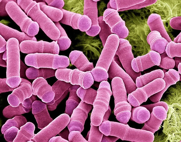
Dividing yeast cells, SEM

 : Dividing yeast cells, SEM
Sold 2
In Stock
We accept all major payment options so you can pay for the things you need in the way that is most convenient for you

Choose Your Product


Science Photo Library Photo Prints and Wall Art

Dividing yeast cells, SEM

Dividing yeast cells. Coloured scanning electron micrograph (SEM) of Schizosaccharomyces pombe yeast cells dividing. S. pombe is a single-celled fungus that is studied widely as a model organism for eukaryotic cell division. It is a rod-shaped yeast that grows by elongation at its ends. It replicates by binary fission. When it reaches a certain size its genetic material (deoxyribonucleic acid, DNA) separates to opposite ends of the cell and a division septum (wall) grows across the centre of the cell, dividing it into two daughter cells that are identical to the parent cell. Magnification: x2400 when printed 10cm wide. Science Photo Library features Science and Medical images including photos and illustrations. © STEVE GSCHMEISSNER/SCIENCE PHOTO LIBRARY

Media ID 6291941

Asexual Binary Fission Dividing Division Eukaryote Eukaryotic Eumycota Fungal Fungi Fungus Magnified Image Microscopic Subjects Model Organism Mycology Naturemycology Re Production Replicating Replication Reproducing Single Celled Yeast Cells False Coloured Micro Biology Microbiological Scanning Electron Microscope

FEATURES IN THESE COLLECTIONS

> Animals > Mammals > Muridae > Sua

> Popular Themes > DNA

> Science > SEM

SECURE PAYMENTS
We accept all major payment options so you can pay for the things you need in the way that is most convenient for you

Made in USA
Safe Shipping, Easy Returns

FREE PERSONALISATION
We are proud to offer a range of customisation including Cropping, Captions, and Filters. Zoom Cropping can be adjusted in the basket


EDITORS COMMENTS

This print showcases the intricate process of dividing yeast cells, captured through a colored scanning electron microscope (SEM). The image depicts Schizosaccharomyces pombe yeast cells in the midst of division, shedding light on the fascinating world of eukaryotic cell division. S. pombe is a rod-shaped single-celled fungus that serves as an invaluable model organism for studying this fundamental biological process. At a magnification of x2400 when printed 10cm wide, the fine details become apparent: elongated yeast cells undergoing binary fission to replicate and reproduce. As these cells grow and reach a certain size, their genetic material (DNA) separates to opposite ends while a division septum gradually forms at the center. This results in two identical daughter cells emerging from what was once a single parent cell. The false-colored representation adds depth and enhances our understanding of this microscopic subject matter. By utilizing SEM technology, scientists are able to delve into the intricacies of cellular biology and gain insights into various microbiological processes. This remarkable image not only highlights the beauty found within nature's smallest creations but also emphasizes how crucial fungi like S. pombe are in advancing our knowledge of reproduction, genetics, and cellular behavior. It serves as a testament to both scientific curiosity and technological innovation in unraveling nature's mysteries at such minuscule scales.


Framed Prints of Dividing yeast cells, SEM

Bring the wonders of the microscopic world into your home or office with Media Storehouse's Framed Prints. This captivating SEM image, titled "Dividing yeast cells," showcases the intricate beauty of Schizosaccharomyces pombe yeast cells dividing, captured in vibrant color through scanning electron microscopy by Science Photo Library. Each print is meticulously framed and ready to hang, making it an excellent addition to any space, inspiring curiosity and conversation.


Photo Prints of Dividing yeast cells, SEM

Discover the intricacies of nature with Media Storehouse's Photographic Prints. Feast your eyes on this captivating Coloured Scanning Electron Micrograph (SEM) of Schizosaccharomyces pombe yeast cells dividing, as captured by Science Photo Library. This image offers a breathtaking glimpse into the microscopic world, showcasing the dividing yeast cells in vibrant colours. An essential addition to any scientific or educational space, this print ignites curiosity and inspires wonder, making it a perfect conversation starter.


Poster Prints of Dividing yeast cells, SEM

"Discover the wonders of the microscopic world with our Media Storehouse Poster Prints. Feast your eyes on the captivating image of Dividing Yeast Cells, captured in stunning detail through Scanning Electron Microscopy (SEM) by Science Photo Library. This print showcases Schizosaccharomyces pombe yeast cells in the process of division, offering a unique glimpse into the intricate beauty of single-celled organisms studied as model systems in scientific research. Bring this vibrant and educational poster into your home or office to inspire curiosity and ignite conversations about the microscopic world around us."


Jigsaw Puzzles of Dividing yeast cells, SEM

Discover the fascinating world of microbiology with Media Storehouse's Jigsaw Puzzles. Our latest addition to the collection showcases the captivating image of "Dividing yeast cells, SEM" by Science Photo Library. Delve into the intricacies of Schizosaccharomyces pombe yeast cells as they divide, brought to life in intricately detailed pieces. A perfect puzzle for science enthusiasts, biology students, or anyone seeking a challenging and educational pastime.

diamond
Made With High Grade Materials
thumb_up
Professional Quality Finish
package_2
Carefully Packed To Aid Safe Arrival

You May Also Like


Spain, Castile La Mancha, Toledo, San Martins Bridge and Tagus RiverSpain, Castile La Mancha, Toledo, San Martins Bridge and Tagus River
Add to Basket
Street scene in Berlin, GermanyStreet scene in Berlin, Germany
Add to Basket
Suffrage Demonstration N. U. W. S. SSuffrage Demonstration N. U. W. S. S
Add to Basket
Walter Butler in One of the Best (1927)Walter Butler in One of the Best (1927)
Add to Basket
Short C-Class flying boat VH-ABD of QantasShort C-Class flying boat VH-ABD of Qantas
Add to Basket
Humanized animals illustrations: RoosterHumanized animals illustrations: Rooster
Add to Basket
Humanized animals illustrations: HareHumanized animals illustrations: Hare
Add to Basket
Humanized animals illustrations: FlowersHumanized animals illustrations: Flowers
Add to Basket
John Hunter, 1786. Artist: WO GellerJohn Hunter, 1786. Artist: WO Geller
Add to Basket
Hastings, 2009. Creator: Ethel DaviesHastings, 2009. Creator: Ethel Davies
Add to Basket
Costa Rica, Sarapiqui River Valley. Strawberry poison dart frog on limb. Credit asCosta Rica, Sarapiqui River Valley. Strawberry poison dart frog on limb. Credit as
Add to Basket
SCHOOL DESEGREGATION, 1965. African American children on their way to school inSCHOOL DESEGREGATION, 1965. African American children on their way to school in
Add to Basket

Gallery Wall Inspiration

Watson and Crick with their DNA model
Rosalind Franklin
DNA molecule
DNA transcription, molecular model
Double-stranded RNA molecule
DNA molecule
Watson and Crick, DNA discovers
DNA molecule, artwork
The Tarbet Hotel, Loch Lomond, Caledonian Railway poster, 1920
Peebles for Health, Rest and Recreation, railway poster, 1900-1922
Computer screen showing a human genetic sequence
X and Y chromosomes
DNA molecule
Mitosis, light micrograph
Rough endoplasmic reticulum, TEM
Cell structure
Herpes virus replicating
Fingerprint scanner
Computer artwork of a beta DNA segment and spheres
Nucleotide base matrix
DNA molecule, computer model
Nucleosome molecule
DNA molecule, abstract image
DNA nucleosome, molecular model
Dividing yeast cells, SEM
E. coli bacterium

Discover Our Collections


SEMSEM
View Collection
DNADNA
View Collection
SuaSua
View Collection


© Copyright Media Storehouse, All Rights Reserved 2003 - 2025
Facebook     Pinterest     100% Payment Secure     Reviews IO