Fraud Blocker Skip to main content

Micro Biology Collection (page 10)

Microbiology is a fascinating field that delves into the intricate world of microscopic organisms

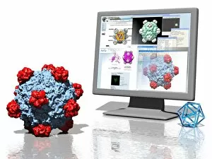
Background imageMicro Biology Collection: Virus research, artwork

Virus research, artwork
Virus research. Conceptual computer artwork representing research into viruses, showing a virus particle (lower left) and a computer screen displaying data on the virus

Background imageMicro Biology Collection: Avian flu research, conceptual artwork

Avian flu research, conceptual artwork
Avian flu research, conceptual computer artwork. Researchers are trying to develop treatments and vaccines to avian flu before it can mutate into a form that can spread between humans

Background imageMicro Biology Collection: Legionella bacteria culture

Legionella bacteria culture
MODEL RELEASED. Legionella bacteria culture. Microbiologist holding a petri dish containing a culture of Legionella sp. bacteria. Legionella sp

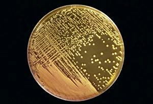
Background imageMicro Biology Collection: Cultured Staphylococcus bacteria

Cultured Staphylococcus bacteria
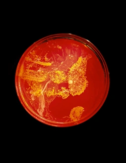
Staphylococcus bacteria. Cultured colonies of Staphylococcus aureus bacteria in a petri dish. S. aureus is a Gram-positive, non- motile

Background imageMicro Biology Collection: Bacillus cereus food poisoning

Bacillus cereus food poisoning
MODEL RELEASED. Bacillus cereus food poisoning. Microbiologist holding a petri dish containing a culture of B. cereus bacteria in one hand and a sample of reheated rice in the other. B

Background imageMicro Biology Collection: Listeria culture

Listeria culture
MODEL RELEASED. Listeria culture. Microbiologist holding a petri dish containing a culture of Listeria monocytogenes bacteria

Background imageMicro Biology Collection: Making money from designer microbes

Making money from designer microbes. Conceptual computer artwork depicting US dollar signs in petri dishes, representing the economic benefits of creating artificial micro-organisms

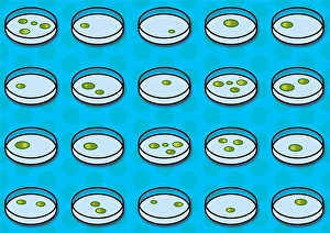
Background imageMicro Biology Collection: Cultures on petri dishes

Cultures on petri dishes
Bacterial cultures on petri dishes. Computer artwork of bacteria growing on petri dishes

Background imageMicro Biology Collection: Bacillus cereus culture

Bacillus cereus culture
MODEL RELEASED. Bacillus cereus culture. Microbiologist holding a petri dish containing a culture of B. cereus bacteria. B

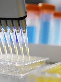
Background imageMicro Biology Collection: Multi-channel pipette

Multi-channel pipette. Multi-channel pipette being used to fill a multi-well sample tray with samples of fluid for analysis

Background imageMicro Biology Collection: Laboratory research

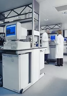
Laboratory research
MODEL RELEASED. Laboratory research. Two researchers talking by a computer in a laboratory

Background imageMicro Biology Collection: Drinking water testing

Drinking water testing
MODEL RELEASED. Drinking water testing. Microbiologist pipetting water onto a petri dish for laboratory analysis. The sample is from an aquifer that supplies drinking water

Background imageMicro Biology Collection: Decomposition

Decomposition
Decompositon. Water vapour rising from decaying organic matter on an active (hot) compost heap. This heat is a by-product of the breakdown of the organic matter by microbes living in the compost heap

Background imageMicro Biology Collection: Severe acute respiratory syndrome (SARS)

Severe acute respiratory syndrome (SARS). Conceptual image of the letters of SARS on a lung artwork. This represents the SARS virus infecting the lungs

Background imageMicro Biology Collection: Macrophage engulfing bacteria, artwork

Macrophage engulfing bacteria, artwork
Macrophage engulfing bacteria. Computer artwork of a macrophage white blood cell (pink) engulfing bacteria (blue). This process is called phagocytosis

Background imageMicro Biology Collection: Neutrophils and Shigella bacteria, SEM

Neutrophils and Shigella bacteria, SEM
Neutrophils and Shigella bacteria. Coloured scanning electron micrograph (SEM) of neutrophil white blood cells (large, yellow-green) and Shigella bacteria (orange, upper right)

Background imageMicro Biology Collection: Macrophages and tuberculosis vaccine

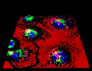
Macrophages and tuberculosis vaccine. Fluorescence light micrograph of macrophage (red, outlined in black) white blood cells that have phagocytosed (engulfed) Mycobacterium bovis bacteria (pink)

Background imageMicro Biology Collection: Neutrophils and anthrax bacteria, SEM

Neutrophils and anthrax bacteria, SEM
Neutrophils and anthrax bacteria. Coloured scanning electron micrograph (SEM) of neutrophil white blood cells (yellow) and Bacillus anthracis bacteria (orange)

Background imageMicro Biology Collection: Neutrophil and trapped bacteria, TEM

Neutrophil and trapped bacteria, TEM
Neutrophil and trapped bacteria. Coloured transmission electron micrograph (TEM) of Neisseria gonorrhoeae bacteria (blue) trapped by a neutrophil (orange), a type of white blood cell

Background imageMicro Biology Collection: Macrophage and tuberculosis vaccine, TEM

Macrophage and tuberculosis vaccine, TEM
Macrophage and tuberculosis vaccine. Coloured transmission electron micrograph (TEM) of phagocytosed (engulfed) Mycobacterium bovis bacteria (purple) in a macrophage (green) white blood cell

Background imageMicro Biology Collection: Macrophage engulfing bacteria, SEM

Macrophage engulfing bacteria, SEM
Macrophage engulfing bacteria. Coloured scanning electron micrograph (SEM) of a macrophage white blood cell (brown) engulfing Mycobacterium tuberculosis bacteria (yellow)

Background imageMicro Biology Collection: Macrophage engulfing tuberculosis vaccine

Macrophage engulfing tuberculosis vaccine. Coloured scanning electron micrograph (SEM) of a macrophage white blood cell engulfing Mycobacterium bovis bacteria (yellow)

Background imageMicro Biology Collection: Neutrophil cell and bacteria, SEM

Neutrophil cell and bacteria, SEM
Neutrophil cell and bacteria. Coloured scanning electron micrograph (SEM) of bacteria (rod-shaped) and part of a neutrophil cell (lower left)

Background imageMicro Biology Collection: Macrophage cell engulfing bacteria, TEM

Macrophage cell engulfing bacteria, TEM
Macrophage cell engulfing bacteria. Coloured transmission electron micrograph (TEM) of bacteria (centre, rod-shaped) inside a macrophage cell

Background imageMicro Biology Collection: Protozoan microscopy, 19th century

Protozoan microscopy, 19th century
Protozoan microscopy. 19th century artwork of microscopic details of protozoan micro-organisms, seen under a microscope. These include a parasitic worm-like organism (Monocystis lumbricorum)

Background imageMicro Biology Collection: Testing for bacteria

Testing for bacteria. Bacteriologist using a strip of growth medium gel to sample bacteria from a chopping board. This is being done to study the number

Background imageMicro Biology Collection: Bacterial colonies

Bacterial colonies. Petri dish containing colonies of bacteria cultured from a hand print found in a house. The print was lifted from a surface on a growth medium gel

Background imageMicro Biology Collection: View of a petri dish with bacterial cultures

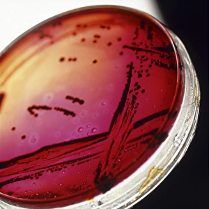
View of a petri dish with bacterial cultures

Background imageMicro Biology Collection: Bacteria water test

Bacteria water testing. Female researcher removing a membrane filter from a bacterial culture dish with forceps as she tests a water sample for bacterial contamination

Background imageMicro Biology Collection: Gloved hand of a researcher holding a petri dish

Gloved hand of a researcher holding a petri dish
Petri dish. Gloved hand of a researcher using forceps to place a grid on an agar plate in order to grow colonies of bacteria as part of a water purity check

Background imageMicro Biology Collection: Technician with bacterial cultures in petri dishes

Technician with bacterial cultures in petri dishes
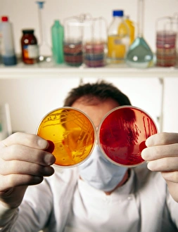
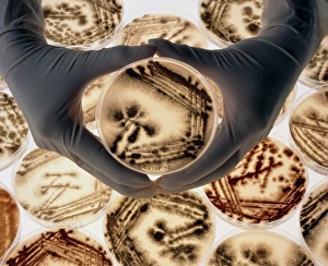
MODEL RELEASED. Bacterial cultures. Masked techni- cian holding two petri dishes containing bacterial cultures in gloved hands

Background imageMicro Biology Collection: Stack of petri dishes with bacterial cultures

Stack of petri dishes with bacterial cultures
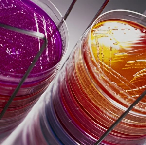
Bacterial cultures. View of a stack of petri dishes containing bacterial cultures. These petri dishes are filled with different coloured agar nutrient media on which bacteria are grown

Background imageMicro Biology Collection: Petri dishes containing bacterial cultures

Petri dishes containing bacterial cultures
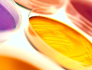
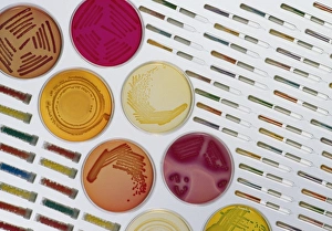
Bacterial culture. View of petri dishes containing bacterial cultures. These petri dishes are filled with different coloured agar nutrient media on which bacteria are grown

Background imageMicro Biology Collection: Close-up of petri dishes with bacterial cultures

Close-up of petri dishes with bacterial cultures
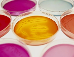
Bacterial culture. Close-up of petri dishes containing bacterial cultures. These petri dishes are filled with different coloured agar nutrient media on which bacteria are grown

Background imageMicro Biology Collection: Hands with petri dishes with Aspergillus cultures

Hands with petri dishes with Aspergillus cultures

Background imageMicro Biology Collection: Researcher testing the sensitivity of bacteria

Researcher testing the sensitivity of bacteria
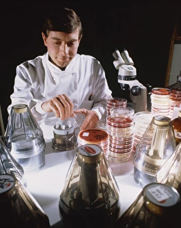
Model released. Researcher testing the sensitivity of bacteria on culture plates. The plunger-like device which he is using is able to dispense six small portions of different antibiotics

Background imageMicro Biology Collection: Quality control on media to grow microorganisms

Quality control on media to grow microorganisms
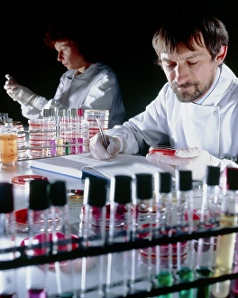
Model released. Researchers testing the performance of different media used to grow microorganisms such as bacteria. The image was taken in one of the 52 laboratories found throughout the UK forming

Background imageMicro Biology Collection: Researcher & samples

Researcher & samples
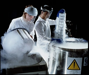
Model released. Researcher removing trays containing samples of bacteria which were stored at low temperature in liquid nitrogen

Background imageMicro Biology Collection: Researchers handling trays of frozen bacteria

Researchers handling trays of frozen bacteria
Model released. Researchers removing trays containing samples of bacteria which were stored at low temperature in liquid nitrogen

Background imageMicro Biology Collection: Different media used to store or grow bacteria

Different media used to store or grow bacteria
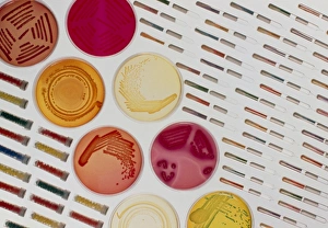
Bacteria in culture. Different media used to grow or store cultures of bacteria. At centre some Petri dishes are seen in which bacteria grow in agar

Background imageMicro Biology Collection: Searching for bacteria in frozen food

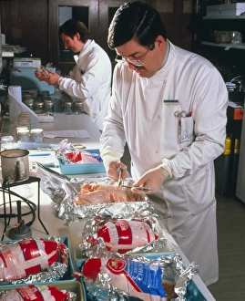
Searching for bacteria in frozen food

Background imageMicro Biology Collection: Media use for growing or storing bacteria

Media use for growing or storing bacteria
Bacteria in culture. Different media used to grow or store cultures of bacteria. At centre some Petri dishes are seen in which bacteria grow in agar

Background imageMicro Biology Collection: Agar filling machine

Agar filling machine
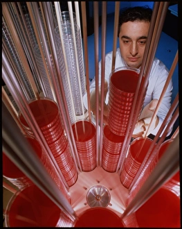
MODEL RELEASED. Agar filling machine. Technician lifts filled petri dishes from an agar machine. Empty petri dishes are at upper left. Agar is a nutrient medium upon which bacteria can be grown

Background imageMicro Biology Collection: AIDS viruses budding from T-cell

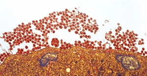
AIDS viruses budding from T-cell
AIDS viruses. Coloured transmission electron micrograph (TEM) of rounded Human Immunodeficiency viruses (HIV) budding from the surface of an infected T-lymphocyte blood cell (T-cell)

Background imageMicro Biology Collection: Adenovirus, TEM

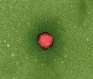
Adenovirus, TEM

Background imageMicro Biology Collection: Vaccinia virus particles, SEM

Vaccinia virus particles, SEM
Vaccinia virus particles. Coloured scanning electron micrograph (SEM) of vaccinia virus particles (red) budding from a cell (blue and green) after replicating

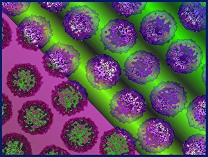
Background imageMicro Biology Collection: Polyoma viruses

Polyoma viruses. Coloured transmission electron micrograph (TEM) of polyoma viruses. These simian (monkey) viruses (SV40) are a type of papovavirus

Background imageMicro Biology Collection: Insect viruses, TEM

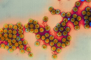
Insect viruses, TEM
Insect viruses. Coloured transmission electron micrograph (TEM) of unidentified isometric insect viruses. These viruses most commonly infect their insect hosts when they feed. Magnification unknown


All Professionally Made to Order for Quick Shipping

Microbiology is a fascinating field that delves into the intricate world of microscopic organisms. From budding yeast cells to calcareous phytoplankton, these tiny beings hold immense importance in our ecosystem. Take a closer look through the lens of a scanning electron microscope (SEM), and you'll be amazed by their intricate structures. One such example is the avian flu virus, which can cause devastating outbreaks among bird populations. Its unique shape and composition are revealed under SEM, highlighting its potential threat to both animals and humans alike. Diatoms, another group of microorganisms, showcase stunning beauty when observed through SEM. Their delicate silica shells form intricate patterns that resemble works of art. These diatoms play a crucial role in marine ecosystems as primary producers. E. coli bacteria, often associated with foodborne illnesses, reveal their rod-shaped structure under SEM. Understanding their morphology helps scientists develop strategies to combat infections caused by this notorious bacterium. The PSCI2A-00015 embryonic stem cell captured under SEM demonstrates its incredible regenerative capabilities. This versatile cell type holds great promise for future medical advancements and treatments. Salmonella bacteria also come into focus under SEM, displaying their flagella-covered surface responsible for motility and infection transmission. Studying these pathogens aids in developing effective prevention measures against salmonellosis. Intriguingly colored TEM images unveil Yersinia pestis bacteria responsible for causing deadly diseases like bubonic plague throughout history. Unveiling their detailed structure allows researchers to better understand how they function within host organisms. Another captivating diatom species reveals itself through SEM imaging - showcasing nature's artistic side once again. These single-celled algae contribute significantly to global carbon fixation while providing habitats for countless aquatic creatures. Candida fungus captures attention with its filamentous appearance when viewed at high magnification using SEM techniques. This opportunistic pathogen can cause various infections in humans ranging from mild oral thrush to life-threatening systemic infections.