Fraud Blocker Skip to main content

Micro Biology Collection (page 4)

Microbiology is a fascinating field that delves into the intricate world of microscopic organisms

Background imageMicro Biology Collection: Sindbis virus capsid protein

Sindbis virus capsid protein, molecular model

Background imageMicro Biology Collection: Tobacco necrosis virus research, artwork

Tobacco necrosis virus research, artwork
Tobacco necrosis virus research, computer artwork

Background imageMicro Biology Collection: Biohazard symbol and virus

Biohazard symbol and virus. Computer artwork of the symbol for a biohazard (red) superimposed on a virus (blue). A biohazard is an organism or biological substance that is harmful to human health

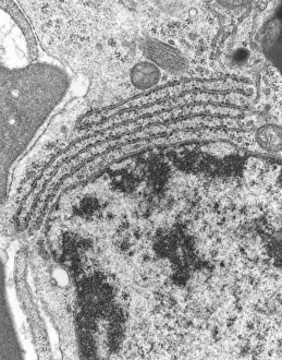
Background imageMicro Biology Collection: TEM of endoplasmic reticulum in mammalian cell

TEM of endoplasmic reticulum in mammalian cell
Rough endoplasmic reticulum. Transmission Electron Micrograph (TEM) of a section through a mammalian cell revealing rough endoplasmic reticulum (ER)

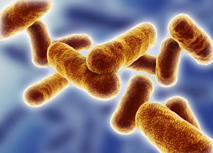
Background imageMicro Biology Collection: Rod shaped bacillus bacteria

Rod shaped bacillus bacteria
3D-computer artwork of bacillus bacteria. These rod-shaped bacteria (bacilli) are widespread in soil and in the air. Many bacillus bacteria are responsible for food spoilage

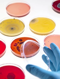
Background imageMicro Biology Collection: Bacteria research

Bacteria research
MODEL RELEASED. Bacteria research. Researcher holding an empty petri dish in front of other dishes containing cultured bacteria (dots)

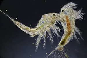
Background imageMicro Biology Collection: Copepods

Copepods. Copepods are microscopic aquatic crustaceans found in waters around the world. They may either be free living or parasitic on other organisms

Background imageMicro Biology Collection: Paramecium protozoa, light micrograph

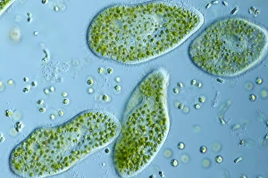
Paramecium protozoa, light micrograph
Paramecium bursaria protozoa, light micrograph. These ciliate protozoa inhabit freshwater, where they feed mainly on bacteria

Background imageMicro Biology Collection: Smallpox virus particle, TEM

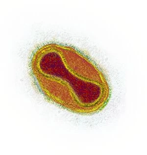
Smallpox virus particle, TEM
Smallpox virus particle. Coloured transmission electron micrograph (TEM) of a Variola major virus particle. Each particle consists of a DNA (deoxyribonucleic acid)

Background imageMicro Biology Collection: Infectious bronchitis virus (IBV), TEM

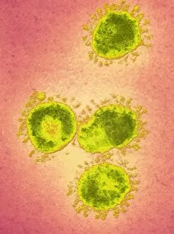
Infectious bronchitis virus (IBV), TEM
Infectious bronchitis virus (IBV) particles, coloured transmission electron micrograph (TEM). This virus is a member of the coronavirus family

Background imageMicro Biology Collection: Eastern equine encephalitis virus, TEM

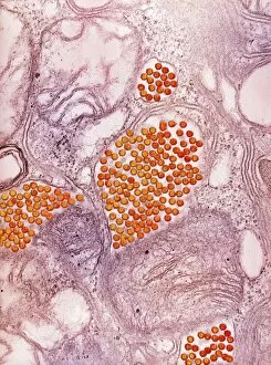
Eastern equine encephalitis virus, TEM
Eastern equine encephalitis virus. Coloured transmission electron micrograph (TEM) of eastern equine encephalitis (EEE) virus particles (Alphavirus sp. orange) in the salivary gland of a mosquito

Background imageMicro Biology Collection: HIV particles, TEM

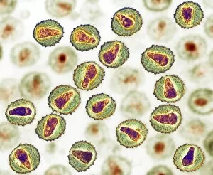
HIV particles, TEM
HIV (human immunodeficiency virus) particles, coloured transmission electron micrograph (TEM). Each particle consists of an RNA (ribonucleic acid, purple/brown) genome

Background imageMicro Biology Collection: Trichodina parasite, light micrograph

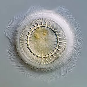
Trichodina parasite, light micrograph
Trichodina parasite. Differential interference contrast micrograph of Trichodina pediculus. This ciliate parasite lives on the skin, gills and mucous membranes of many aquatic animals

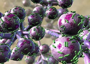
Background imageMicro Biology Collection: Bacterial lung infection

Bacterial lung infection, computer artwork. Rod-shaped bacteria (bacilli, green) on the surface of the alveoli of the lungs (purple)

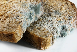
Background imageMicro Biology Collection: Mouldy bread

Mouldy bread. The white areas are fungal mycelia, masses of thread-like structures (hyphae) that absorb nutrients. The blue-green areas are the fungal spores (reproductive structures)

Background imageMicro Biology Collection: Love, microbial art

Love, microbial art. Petri dish containing the fungi Aureobasidium pullulans and Rhodotorula sp. (red). The fungi have been cultured on agar jelly to depict a heart and arrow

Background imageMicro Biology Collection: Bacillus subtilis bacteria, SEM

Bacillus subtilis bacteria, SEM
Bacillus subtilis. Coloured scanning electron micrograph (SEM) of Bacillus subtilis bacteria. B. subtilis, or hay bacillus, is an aerobic, gram-positive bacillus(rod-shaped bacterium)

Background imageMicro Biology Collection: Lactobacillus bacteria, SEM

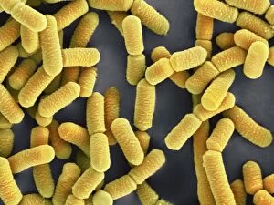
Lactobacillus bacteria, SEM
Lactobacillus sp. bacteria, coloured scanning electron micrograph (SEM). Lactobacillus bacteria are probiotic bacteria, which are useful for human and animal health as they produce bacteriocins

Background imageMicro Biology Collection: Clostridium perfringens bacterium with spore

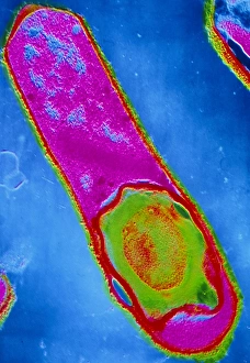
Clostridium perfringens bacterium with spore
False-colour transmission electron micrograph (TEM) of a Clostridium perfringens bacterium with endospore. This species of Gram-positive

Background imageMicro Biology Collection: Bacteria on lung hairs

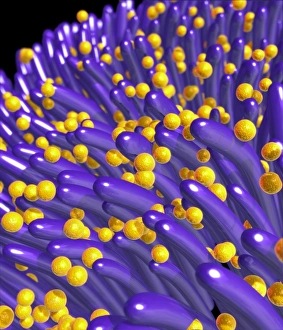
Bacteria on lung hairs
Bacteria in a lung. Computer artwork of bacteria (yellow) on cilia (hair-like structures) in a lung. Bacterial infection of the lungs can cause potentially fatal pneumonia

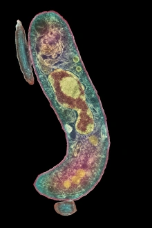
Background imageMicro Biology Collection: Proteus mirabilis bacterium

Proteus mirabilis bacterium
False-colour transmission electron micrograph of the bacterium Proteus mirabilis. It is an enterobacterium which is present normally in the human intestine

Background imageMicro Biology Collection: Spirochete bacteria, TEM

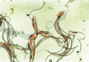
Spirochete bacteria, TEM
Spirochete bacteria, coloured transmission electron micrograph (TEM). Spirochetes are bacteria with helically coiled cells

Background imageMicro Biology Collection: Coloured SEM of Clostridium botulinum

Coloured SEM of Clostridium botulinum
Food poisoning bacterium. Coloured scanning electron micrograph of the Gram-positive anaerobic bacteria Clostridium botulinum, responsible for serious food poisoning in humans. C

Background imageMicro Biology Collection: Rod-shaped bacteria

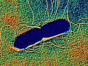
Rod-shaped bacteria. Computer artwork of rod- shaped bacteria (bacilli). Typical bacilli are E. coli and Salmonella bacteria, though there are many others as well

Background imageMicro Biology Collection: Root nodule

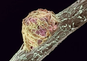
Root nodule. Coloured scanning electron micrograph (SEM) of a root nodule on a pea plant (Pisum sativum) caused by the nitrogen-fixing soil bacteria Rhizobium leguminosarum

Background imageMicro Biology Collection: Yeast cells, TEM

Yeast cells, TEM
Yeast cells, coloured transmission electron micrograph (TEM). The cell wall is shown in blue, cytoplasm in red, and vacuole in green. Yeast is a single-celled fungus that reproduces asexually

Background imageMicro Biology Collection: PSCI2A-00079

PSCI2A-00079
Louis Pasteur supervising an inoculation for hydrophobia (rabies), 1880s. Hand-colored woodcut of a 19th-century photograph

Background imageMicro Biology Collection: PASTEUR, Louis (1822-1895)

PASTEUR, Louis (1822-1895). French chemist and bacteriologist, pioneer of the microbiology. Painting

Background imageMicro Biology Collection: Engraving of Louis Pasteur French chemist and bacteriologist

Engraving of Louis Pasteur French chemist and bacteriologist
PASTEUR, Louis (1822-1895) French chemist and bacteriologist. Study of microbes in the Pasteur Institute. Engraving

Background imageMicro Biology Collection: Ciliate protozoa, light micrograph

Ciliate protozoa, light micrograph
Ciliate protozoa. Light micrograph of a mixed population of freshwater ciliate protozoa and algae. The algae are the green filaments crossing the field

Background imageMicro Biology Collection: Orbulina foraminiferan, SEM

Orbulina foraminiferan, SEM
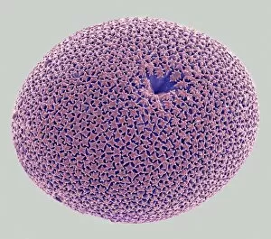
Orbulina. Coloured scanning electron micrograph (SEM) of the shell of the foraminiferan Orbulina sp. Foraminiferans are marine single-celled protists that construct and inhabit shells (tests)

Background imageMicro Biology Collection: Ciliate protozoa dividing

Ciliate protozoa dividing
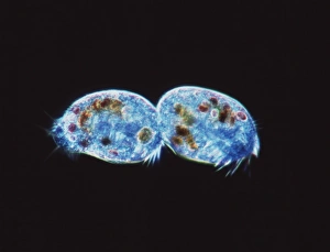
Ciliate protozoa. Light micrograph of unidentified ciliate protozoa (class Ciliata) undergoing asexual reproduction. These unicellular micro-organisms can multiply by either of two methods

Background imageMicro Biology Collection: Plankton cell wall, SEM

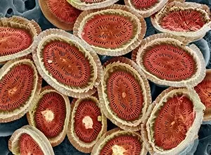
Plankton cell wall, SEM
Plankton cell wall. Coloured scanning electron micrograph (SEM) of the geometrical plates making up the mineralised cell wall of a planktonic alga (Coronosphaera mediterranea)

Background imageMicro Biology Collection: Copepod crustacean, light micrograph

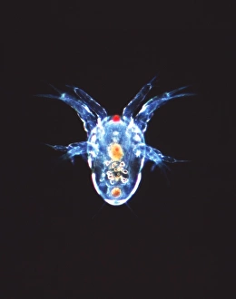
Copepod crustacean, light micrograph
Copepod crustacean. Light micrograph of the nauplius larva of a copepod crustacean (subclass Copepoda). This is the first stage in the life cycle of a copepod

Background imageMicro Biology Collection: Euglena protozoan, SEM

Euglena protozoan, SEM
Euglena protozoan (Euglena sp.), coloured scanning electron micrograph (SEM). This protozoan is described as flagellate because it has a flagellum (thin tail-like structure)

Background imageMicro Biology Collection: Ciliate protozoans, SEM

Ciliate protozoans, SEM
Ciliate protozoans. Coloured scanning electron micrograph (SEM) of sessile (anchored) ciliate protozoan (subclass Peritrichia)

Background imageMicro Biology Collection: Giardia protozoan, TEM

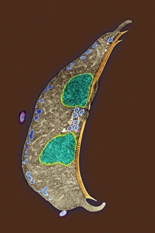
Giardia protozoan, TEM
Giardia protozoan. Coloured transmission electron micrograph (TEM) of a Giardia sp. protozoan. It is a single-celled organism that has two cell nuclei (green)

Background imageMicro Biology Collection: Rotifer worm, light micrograph

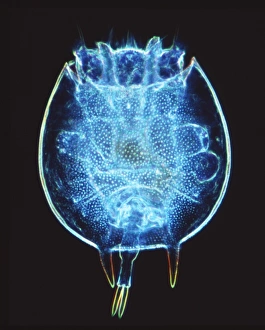
Rotifer worm, light micrograph
Rotifer worm. Light micrograph of the rotifer worm Platyas quadricornis. This free-swimming, aquatic micro-organism has a body which is divided into three sections

Background imageMicro Biology Collection: Ciliate protozoa, SEM

Ciliate protozoa, SEM
Ciliate protozoa. Coloured scanning electron micrograph (SEM) of cilliate protozoa (unicellular animals). The ciliates are so named because of the hair-like cilia surrounding their bodies which are

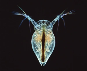
Background imageMicro Biology Collection: Water flea, light micrograph

Water flea, light micrograph
Water flea. Light micrograph of a water flea (suborder Cladocera). The long antennae (upper left and right) of this freshwater crustacean have swimming hairs and serve as rowing organs

Background imageMicro Biology Collection: Flagellate protozoan, SEM

Flagellate protozoan, SEM
Flagellate protozoan, coloured scanning electron micrograph (SEM). This is a protozoan that has a flagellum (thin tail-like structure, lower left)

Background imageMicro Biology Collection: Parasitic protozoan, TEM

Parasitic protozoan, TEM
Parasitic protozoan. Coloured transmission electron micrograph (TEM) of a trypanosome protozoan (Trypanosoma sp.). The nucleus (centre), cell membrane (outline)

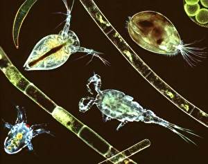
Background imageMicro Biology Collection: Aquatic crustaceans, light micrograph

Aquatic crustaceans, light micrograph
Aquatic crustaceans. Composite image of light micrographs of aquatic planktonic crustaceans with strands of filamentous algae

Background imageMicro Biology Collection: Lychnocanium radiolarian, SEM

Lychnocanium radiolarian, SEM
Lychnocanium radiolarian. Coloured scanning electron micrograph (SEM) of the shell of a Lychnocanium sp. radiolarian. Radiolaria are single-celled protozoans that are found in marine plankton

Background imageMicro Biology Collection: Alga-covered protozoan, SEM

Alga-covered protozoan, SEM
Alga-covered protozoan, coloured scanning electron micrograph (SEM). Algae (blue) are seen on the body of the protozoan, and cilia (green) are used to waft the algae to the protozoans feeding area

Background imageMicro Biology Collection: Paramecium protozoa, SEM

Paramecium protozoa, SEM
Paramecium protozoa (Paramecium sp.), coloured scanning electron micrograph (SEM). These protozoa (two seen) are described as ciliate because many cilia (hair-like structures) cover them

Background imageMicro Biology Collection: Hartmannella amoeba and bacterium

Hartmannella amoeba and bacterium. Computer artwork of a Hartmannella vermiformis amoeba (green) engulfing a Legionella pneumophila bacterium (orange) with a pseudopodium

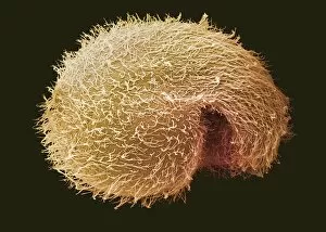
Background imageMicro Biology Collection: Breslauides ciliate protozoan, SEM

Breslauides ciliate protozoan, SEM
Breslauides ciliate protozoan. Coloured scanning electron micrograph (SEM) of a ciliate protozoan (Breslauides sp.). This tiny single-celled organism is found in water


All Professionally Made to Order for Quick Shipping

Microbiology is a fascinating field that delves into the intricate world of microscopic organisms. From budding yeast cells to calcareous phytoplankton, these tiny beings hold immense importance in our ecosystem. Take a closer look through the lens of a scanning electron microscope (SEM), and you'll be amazed by their intricate structures. One such example is the avian flu virus, which can cause devastating outbreaks among bird populations. Its unique shape and composition are revealed under SEM, highlighting its potential threat to both animals and humans alike. Diatoms, another group of microorganisms, showcase stunning beauty when observed through SEM. Their delicate silica shells form intricate patterns that resemble works of art. These diatoms play a crucial role in marine ecosystems as primary producers. E. coli bacteria, often associated with foodborne illnesses, reveal their rod-shaped structure under SEM. Understanding their morphology helps scientists develop strategies to combat infections caused by this notorious bacterium. The PSCI2A-00015 embryonic stem cell captured under SEM demonstrates its incredible regenerative capabilities. This versatile cell type holds great promise for future medical advancements and treatments. Salmonella bacteria also come into focus under SEM, displaying their flagella-covered surface responsible for motility and infection transmission. Studying these pathogens aids in developing effective prevention measures against salmonellosis. Intriguingly colored TEM images unveil Yersinia pestis bacteria responsible for causing deadly diseases like bubonic plague throughout history. Unveiling their detailed structure allows researchers to better understand how they function within host organisms. Another captivating diatom species reveals itself through SEM imaging - showcasing nature's artistic side once again. These single-celled algae contribute significantly to global carbon fixation while providing habitats for countless aquatic creatures. Candida fungus captures attention with its filamentous appearance when viewed at high magnification using SEM techniques. This opportunistic pathogen can cause various infections in humans ranging from mild oral thrush to life-threatening systemic infections.