Fraud Blocker Skip to main content

Micro Biology Collection (page 59)

Microbiology is a fascinating field that delves into the intricate world of microscopic organisms

Background imageMicro Biology Collection: Virus particles, conceptual artwork

Virus particles, conceptual artwork
Virus particles, conceptual computer artwork. Viruses contain a core of genetic material, either DNA (deoxyribonucleic acid) or RNA (ribonucleic acid) surrounded by a protein coat, or capsid

Background imageMicro Biology Collection: Poliovirus particles, TEM

Poliovirus particles, TEM
Poliovirus particles. Coloured transmissionelectron micrograph (TEM) of poliovirus particles.Each virus consists of a protein coat (capsid)surrounding a core of RNA (ribonucleicacid)

Background imageMicro Biology Collection: Myxococcus xanthus bacteria, SEM

Myxococcus xanthus bacteria, SEM
Myxococcus xanthus bacteria, coloured scanningelectron micrograph (SEM). M. xanthus is a rodshaped Gram-negative bacterium that resides inthe soil

Background imageMicro Biology Collection: Proteus vulgaris bacterium, TEM

Proteus vulgaris bacterium, TEM
Proteus vulgaris bacterium, colouredtransmission electron micrograph (TEM). The yellowthreads are flagella, hair-like structures thatthe bacterium beats for motility

Background imageMicro Biology Collection: Staphylococcus aureus dividing, TEM

Staphylococcus aureus dividing, TEM
Staphylococcus aureus bacteria dividing, coloured transmission electron micrograph. Atlower left is a single bacterium, the bacterium atupper centre is starting to divide

Background imageMicro Biology Collection: Oral bacteria, TEM

Oral bacteria, TEM
Oral bacteria. Computer-enhanced colouredtransmission electron micrograph (TEM) of Porphyromonas gingivalis bacteria (formerlynamed Bacteroides gingivalis )

Background imageMicro Biology Collection: Bacterial cell

Bacterial cell, computer artwork. This is ageneralised bacterium, illustrating features ofdifferent bacterial types that would not becombined in a single cell

Background imageMicro Biology Collection: Helicobacter pylori bacteria in stomach

Helicobacter pylori bacteria in stomach
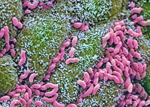
Helicobacter pylori bacteria. Colouredscanning electron micrograph (SEM) of Helicobacter pylori bacteria (pink), a causeof gastritis (stomach lining inflammation)

Background imageMicro Biology Collection: Listeria monocytogenes bacteria, TEM

Listeria monocytogenes bacteria, TEM
Listeria bacteria. Coloured transmissionelectron micrograph (TEM) of a Listeria monocytogenes bacterium (centre to bottomright). It is dividing by a process of unequalcell growth known as budding

Background imageMicro Biology Collection: Unidentified rod-shaped bacteria

Unidentified rod-shaped bacteria
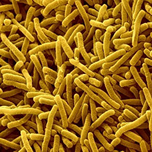
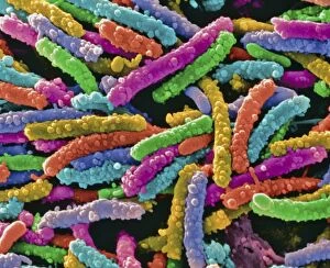
Bacteria. Coloured scanning electronmicrograph (SEM) of unidentified bacilli(rod-shaped bacteria). Magnification unknown

Background imageMicro Biology Collection: Louis Pasteur, French chemist and microbiologist

Louis Pasteur, French chemist and microbiologist. Best known for his scientific breakthroughs in the causes and prevention of diseases


All Professionally Made to Order for Quick Shipping

Microbiology is a fascinating field that delves into the intricate world of microscopic organisms. From budding yeast cells to calcareous phytoplankton, these tiny beings hold immense importance in our ecosystem. Take a closer look through the lens of a scanning electron microscope (SEM), and you'll be amazed by their intricate structures. One such example is the avian flu virus, which can cause devastating outbreaks among bird populations. Its unique shape and composition are revealed under SEM, highlighting its potential threat to both animals and humans alike. Diatoms, another group of microorganisms, showcase stunning beauty when observed through SEM. Their delicate silica shells form intricate patterns that resemble works of art. These diatoms play a crucial role in marine ecosystems as primary producers. E. coli bacteria, often associated with foodborne illnesses, reveal their rod-shaped structure under SEM. Understanding their morphology helps scientists develop strategies to combat infections caused by this notorious bacterium. The PSCI2A-00015 embryonic stem cell captured under SEM demonstrates its incredible regenerative capabilities. This versatile cell type holds great promise for future medical advancements and treatments. Salmonella bacteria also come into focus under SEM, displaying their flagella-covered surface responsible for motility and infection transmission. Studying these pathogens aids in developing effective prevention measures against salmonellosis. Intriguingly colored TEM images unveil Yersinia pestis bacteria responsible for causing deadly diseases like bubonic plague throughout history. Unveiling their detailed structure allows researchers to better understand how they function within host organisms. Another captivating diatom species reveals itself through SEM imaging - showcasing nature's artistic side once again. These single-celled algae contribute significantly to global carbon fixation while providing habitats for countless aquatic creatures. Candida fungus captures attention with its filamentous appearance when viewed at high magnification using SEM techniques. This opportunistic pathogen can cause various infections in humans ranging from mild oral thrush to life-threatening systemic infections.