Fraud Blocker Skip to main content

Central Nervous System Collection (page 7)

The central nervous system, the intricate network that governs our every thought and movement, is a marvel of complexity

Background imageCentral Nervous System Collection: Inflamed brain, conceptual artwork

Inflamed brain, conceptual artwork
Inflamed brain, conceptual computer artwork

Background imageCentral Nervous System Collection: Gamma-aminobutyric acid GABA molecule

Gamma-aminobutyric acid GABA molecule
Gamma-aminobutyric acid (GABA), molecular model. Main inhibitory neurotransmitter in the central nervous system of mammalians

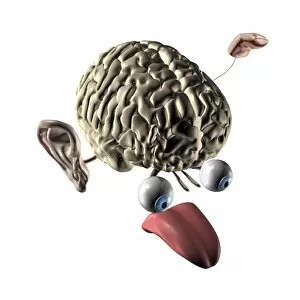
Background imageCentral Nervous System Collection: Human brain and senses, artwork

Human brain and senses, artwork
Human brain and senses. Artwork of a human brain, seen obliquely from the front, with the five senses shown. These are the visual organs (vision, the eyes), the olfactory organ (smell, the nose)

Background imageCentral Nervous System Collection: Brain activity, artwork C017 / 7401

Brain activity, artwork C017 / 7401
Brain activity, computer artwork

Background imageCentral Nervous System Collection: Psychic brain, conceptual image

Psychic brain, conceptual image. Artwork showing the human brain and head, surrounded in flames, with the pineal gland highlighted (bright dot in centre of forehead)

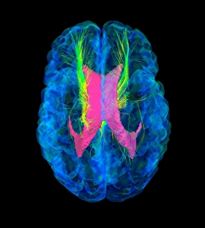
Background imageCentral Nervous System Collection: Brain fibres, DTI MRI scan C017 / 7037

Brain fibres, DTI MRI scan C017 / 7037
Brain fibres. 3D diffusion tensor imaging (DTI) magnetic resonance imaging (MRI) scan of a selection of nerve pathways (green/yellow) in the brain

Background imageCentral Nervous System Collection: Brain fibres, DTI MRI scan C017 / 7038

Brain fibres, DTI MRI scan C017 / 7038
Brain fibres. 3D diffusion tensor imaging (DTI) magnetic resonance imaging (MRI) scan of a selection of nerve pathways (green/yellow) in the brain

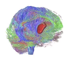
Background imageCentral Nervous System Collection: Brain tumour, DTI MRI scan C017 / 7057

Brain tumour, DTI MRI scan C017 / 7057
Brain tumour. 3D diffusion tensor imaging (DTI) magnetic resonance imaging (MRI) scan of nerve pathways (coloured) in a brain with a tumour (red, centre right)

Background imageCentral Nervous System Collection: Brain fibres, DTI MRI scan C017 / 7100

Brain fibres, DTI MRI scan C017 / 7100
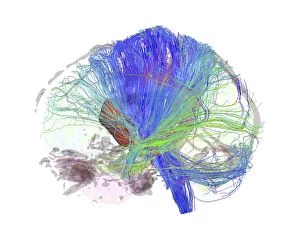
Brain fibres. 3D diffusion tensor imaging (DTI) magnetic resonance imaging (MRI) scan of a selection of nerve pathways (blue, pink, green) in the brain. The front of the brain is at left

Background imageCentral Nervous System Collection: Nerve cells, computer artwork F007 / 1988

Nerve cells, computer artwork F007 / 1988
Nerve cells. Computer artwork of nerve cells, or neurons

Background imageCentral Nervous System Collection: Brain tumour, DTI MRI scan C017 / 7059

Brain tumour, DTI MRI scan C017 / 7059
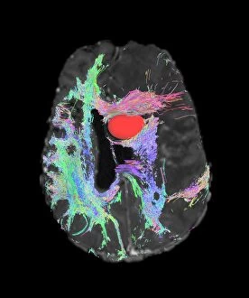
Brain tumour. Axial diffusion tensor imaging (DTI) magnetic resonance imaging (MRI) scan of nerve pathways (coloured) in a brain with a tumour (red, upper centre)

Background imageCentral Nervous System Collection: Brain tumour, DTI MRI scan C017 / 7058

Brain tumour, DTI MRI scan C017 / 7058
Brain tumour. 3D diffusion tensor imaging (DTI) magnetic resonance imaging (MRI) scan of nerve pathways (coloured) in a brain with a tumour (red, centre left)

Background imageCentral Nervous System Collection: Brain, neural network F007 / 0946

Brain, neural network F007 / 0946
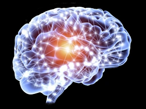
Neural network. Computer artwork of a brain in side view, with the brains neural network represented by lines and flashes. A neural network is made up of nerve cells (neurons)

Background imageCentral Nervous System Collection: Central nervous system, artwork C017 / 7400

Central nervous system, artwork C017 / 7400
Central nervous system, computer artwork

Background imageCentral Nervous System Collection: Pyramidal nerve cell, artwork C017 / 2275

Pyramidal nerve cell, artwork C017 / 2275
Pyramidal nerve cell. Computer artwork of a pyramidal nerve cell from the cerebral cortex of the brain. Pyramidal cells are so named for their triangular cell bodies (lower frame)

Background imageCentral Nervous System Collection: Brain nerve cells, TEM C014 / 0357

Brain nerve cells, TEM C014 / 0357
Brain nerve cells. Transmission electron micrograph (TEM) of a section through brain tissue from the cerebral cortex, showing numerous neurons (nerve cells) surrounded by axons and dendrites

Background imageCentral Nervous System Collection: Neural cells F006 / 9203

Neural cells F006 / 9203
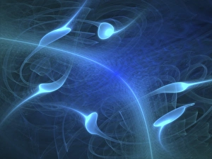
Computer artwork depicting nerve cells and neural communication

Background imageCentral Nervous System Collection: Effect of Parkinsons disease, artwork

Effect of Parkinsons disease, artwork
Effect of Parkinsons disease. Computer artwork showing the neural pathways within the brain that are affected by a lack of dopamine, caused by the onset of Parkinsons disease

Background imageCentral Nervous System Collection: Spondylodiscitis, MRI scan

Spondylodiscitis, MRI scan
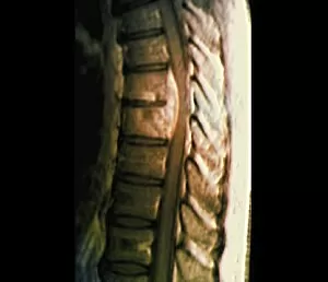
Spondylodiscitis. Coloured magnetic resonance imaging (MRI) scan of a sagittal section through the spine of a 69-year-old patient with spondylodiscitis at the level of the T7-T8 vertebra

Background imageCentral Nervous System Collection: Nerve ganglion, light micrograph

Nerve ganglion, light micrograph
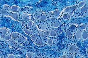
Nerve ganglion. Light micrograph of a section through a dorsal (sensory) spinal root ganglion associated with a sensory nerve root of the spinal cord. Sensory information from peripheral sites e.g

Background imageCentral Nervous System Collection: Brain synapse, anatomical artwork

Brain synapse, anatomical artwork
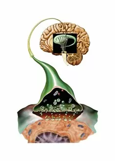
Brain synapse. Anatomical computer artwork of a human brain with an enlargement showing the structure of a synapse (lower right) within one of the striate bodies that make up the striatum

Background imageCentral Nervous System Collection: Spina bifida, MRI scan

Spina bifida, MRI scan
Fibrolipoma of the spine. Magnetic resonance imaging (MRI) scan of a sagittal section through the spine of a 24-month-old patient with spina bifida

Background imageCentral Nervous System Collection: Striatum, anatomical artwork

Striatum, anatomical artwork
Striatum. Anatomical computer artwork of a human brain, showing the striate bodies (blue and green) in the midbrain, and the substantia nigra (black) in the brainstem

Background imageCentral Nervous System Collection: Nerve cell degeneration, artwork

Nerve cell degeneration, artwork
Nerve cell degeneration. Computer artwork showing a collection of nerve cells (neurons) that have become damaged

Background imageCentral Nervous System Collection: Limbic system of the brain, artwork C015 / 4529

Limbic system of the brain, artwork C015 / 4529
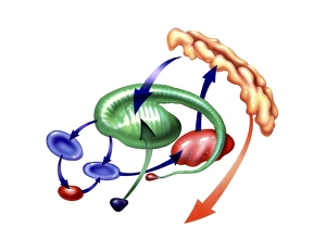
Limbic system of the brain. Computer artwork showing the main parts of the limbic system from the human brain. At centre is the oval-shaped thalamus (red) which receives sensory stimuli

Background imageCentral Nervous System Collection: Hearing centre, artwork

Hearing centre, artwork
Hearing centre. Computer artwork showing the area of the brains temporal lobe that is associated with hearing (red). This area receives auditory input from the cochlea (coiled)
Background imageCentral Nervous System Collection: Human brain, anatomical artwork

Human brain, anatomical artwork
Human brain. Anatomical computer artwork of a human brain seen from below, showing the cranial nerves (centre)

Background imageCentral Nervous System Collection: Fibrolipoma of the spine, MRI scan

Fibrolipoma of the spine, MRI scan
Fibrolipoma of the spine. Coloured magnetic resonance imaging (MRI) scan of a sagittal section through the thoraco-lumbar spine of a 19-month-old patient

Background imageCentral Nervous System Collection: Substantia nigra, artwork

Substantia nigra, artwork
Substantia nigra. Computer artwork showing a healthy substantia nigra (left) and degenerated substantia nigra (right) from human brains

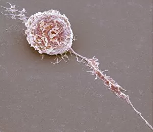
Background imageCentral Nervous System Collection: Microglial white blood cell, SEM C016 / 9115

Microglial white blood cell, SEM C016 / 9115
Microglial white blood cell. Coloured scanning electron micrograph (SEM) of a microglial cell. Microglia are phagocytic macrophage white blood cells that are the main form of immune system in

Background imageCentral Nervous System Collection: Fridtjof Nansen, Norwegian explorer C016 / 8812

Fridtjof Nansen, Norwegian explorer C016 / 8812
Fridtjof Nansen (1861-1930), Norwegian explorer, biologist and humanitarian. In 1888 he was the first to cross the Greenland ice sheet and showed that it covered the entire island

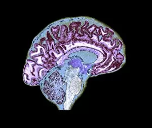
Background imageCentral Nervous System Collection: Normal human brain, MRI scan C016 / 8840

Normal human brain, MRI scan C016 / 8840
Brain. Coloured magnetic resonance imaging (MRI) scan of a sagittal section through a healthy human brain. The highly folded area (yellow) is the cerebrum, which is responsible for conscious thought

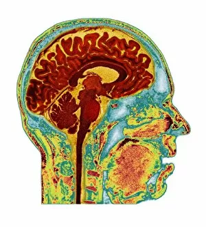
Background imageCentral Nervous System Collection: Normal human brain, MRI scan C016 / 8848

Normal human brain, MRI scan C016 / 8848
Brain. Coloured magnetic resonance imaging (MRI) scan of a sagittal section through a patients head showing a healthy human brain

Background imageCentral Nervous System Collection: Eastern equine encephalitis, TEM C016 / 9390

Eastern equine encephalitis, TEM C016 / 9390
Eastern equine encephalitis. Transmission electron micrograph (TEM) of a section through tissue from a human central nervous system (CNS)

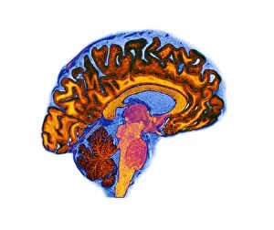
Background imageCentral Nervous System Collection: Normal human brain, MRI scan C016 / 8837

Normal human brain, MRI scan C016 / 8837
Brain. Coloured magnetic resonance imaging (MRI) scan of a sagittal section through a healthy human brain. The highly folded area (orange) is the cerebrum, which is responsible for conscious thought

Background imageCentral Nervous System Collection: Microglial white blood cell, SEM C016 / 9119

Microglial white blood cell, SEM C016 / 9119
Microglial white blood cell. Coloured scanning electron micrograph (SEM) of a microglial cell. Microglia are phagocytic macrophage white blood cells that are the main form of immune system in

Background imageCentral Nervous System Collection: Microglial white blood cell, SEM C016 / 9117

Microglial white blood cell, SEM C016 / 9117
Microglial white blood cell. Coloured scanning electron micrograph (SEM) of a microglial cell. Microglia are phagocytic macrophage white blood cells that are the main form of immune system in

Background imageCentral Nervous System Collection: Normal human brain, MRI scan C016 / 8841

Normal human brain, MRI scan C016 / 8841
Brain. Coloured magnetic resonance imaging (MRI) scan of a sagittal section through a healthy human brain. The highly folded area (red and yellow) is the cerebrum

Background imageCentral Nervous System Collection: Microglial white blood cell, SEM C016 / 9109

Microglial white blood cell, SEM C016 / 9109
Microglial white blood cell. Coloured scanning electron micrograph (SEM) of a microglial cell. Microglia are phagocytic macrophage white blood cells that are the main form of immune system in

Background imageCentral Nervous System Collection: Normal human brain, MRI scan C016 / 8835

Normal human brain, MRI scan C016 / 8835
Brain. Coloured magnetic resonance imaging (MRI) scan of a sagittal section through a healthy human brain. The highly folded area across top is the cerebrum

Background imageCentral Nervous System Collection: Normal human brain, MRI scan C016 / 8846

Normal human brain, MRI scan C016 / 8846
Brain. Coloured magnetic resonance imaging (MRI) scan of a sagittal section through a patients head showing a healthy human brain

Background imageCentral Nervous System Collection: Normal human brain, MRI scan C016 / 8834

Normal human brain, MRI scan C016 / 8834
Brain. Coloured magnetic resonance imaging (MRI) scan of a sagittal section through a healthy human brain. The highly folded area (orange) is the cerebrum, which is responsible for conscious thought

Background imageCentral Nervous System Collection: Normal human brain, MRI scan C016 / 8849

Normal human brain, MRI scan C016 / 8849
Brain. Coloured magnetic resonance imaging (MRI) scan of a sagittal section through a patients head showing a healthy human brain

Background imageCentral Nervous System Collection: Normal human brain, MRI scan C016 / 8836

Normal human brain, MRI scan C016 / 8836
Brain. Coloured magnetic resonance imaging (MRI) scan of a sagittal section through a healthy human brain. The highly folded area across top is the cerebrum

Background imageCentral Nervous System Collection: Normal human brain, MRI scan C016 / 8844

Normal human brain, MRI scan C016 / 8844
Brain. Coloured magnetic resonance imaging (MRI) scan of a sagittal section through a patients head showing a healthy human brain

Background imageCentral Nervous System Collection: Normal human brain, MRI scan C016 / 8839

Normal human brain, MRI scan C016 / 8839
Brain. Coloured magnetic resonance imaging (MRI) scan of a sagittal section through a healthy human brain. The highly folded area across top is the cerebrum


All Professionally Made to Order for Quick Shipping

The central nervous system, the intricate network that governs our every thought and movement, is a marvel of complexity. From the delicate cerebellum tissue to the detailed light micrograph capturing its essence, we are reminded of its importance in maintaining balance and coordination. Anatomy comes alive as we explore the human brain from an inferior view. The intricacy of brain fibers is revealed through DTI MRI scans like C017/7099 and C017/7035, showcasing their vital role in transmitting information throughout this extraordinary organ. Artistic renderings bring us closer to understanding the medulla oblongata's significance within the brain. Its portrayal in various artworks allows us to appreciate how it controls essential functions such as breathing and heart rate. As we delve deeper into studying the central nervous system, models of the human brain provide invaluable insights into its structure and organization. Lateral views reveal countless regions responsible for cognition, emotion regulation, sensory perception, and motor control. Microscope slides offer glimpses into nerve cells' intricate architecture—a testament to their ability to transmit electrical signals at lightning speed. Meanwhile, glial stem cell cultures captured under a light microscope remind us of their crucial role in supporting neuronal function. Finally, artistic representations unveil the limbic system's enigmatic nature—an interconnected web responsible for emotions and memory formation. These captivating artworks allow us to visualize this complex network within our brains. Exploring these hints provides a glimpse into the awe-inspiring world of our central nervous system—the very foundation upon which our thoughts, actions, memories reside—reminding us just how remarkable our brains truly are.