Fraud Blocker Skip to main content

Central Nervous System Collection (page 9)

The central nervous system, the intricate network that governs our every thought and movement, is a marvel of complexity

Background imageCentral Nervous System Collection: Microglial white blood cell, SEM C016 / 9114

Microglial white blood cell, SEM C016 / 9114
Microglial white blood cell. Coloured scanning electron micrograph (SEM) of a microglial cell. Microglia are phagocytic macrophage white blood cells that are the main form of immune system in

Background imageCentral Nervous System Collection: Microglial white blood cells, SEM C016 / 9113

Microglial white blood cells, SEM C016 / 9113
Microglial white blood cells. Coloured scanning electron micrograph (SEM) of microglial cells. Microglia are phagocytic macrophage white blood cells that are the main form of immune system in

Background imageCentral Nervous System Collection: Synapse, artwork C016 / 7535

Synapse, artwork C016 / 7535
Synapse. Computer artwork of a synapse, the junction between nerve cells (orange). Synapses transmit electrical signals from one nerve cell to the next
Background imageCentral Nervous System Collection: Microglial white blood cell, SEM C016 / 9118

Microglial white blood cell, SEM C016 / 9118
Microglial white blood cell. Coloured scanning electron micrograph (SEM) of a microglial cell. Microglia are phagocytic macrophage white blood cells that are the main form of immune system in

Background imageCentral Nervous System Collection: Microglial white blood cells, SEM C016 / 9111

Microglial white blood cells, SEM C016 / 9111
Microglial white blood cells. Coloured scanning electron micrograph (SEM) of microglial cells. Microglia are phagocytic macrophage white blood cells that are the main form of immune system in

Background imageCentral Nervous System Collection: Normal human brain, MRI scan C016 / 8847

Normal human brain, MRI scan C016 / 8847
Brain. Coloured magnetic resonance imaging (MRI) scan of a sagittal section through a patients head showing a healthy human brain

Background imageCentral Nervous System Collection: Microglial white blood cells, SEM C016 / 9112

Microglial white blood cells, SEM C016 / 9112
Microglial white blood cells. Coloured scanning electron micrograph (SEM) of microglial cells. Microglia are phagocytic macrophage white blood cells that are the main form of immune system in

Background imageCentral Nervous System Collection: Microglial white blood cell, SEM C016 / 9116

Microglial white blood cell, SEM C016 / 9116
Microglial white blood cell. Coloured scanning electron micrograph (SEM) of a microglial cell. Microglia are phagocytic macrophage white blood cells that are the main form of immune system in

Background imageCentral Nervous System Collection: Human brain, artwork C016 / 9370

Human brain, artwork C016 / 9370
Human brain, computer artwork

Background imageCentral Nervous System Collection: Parkinsons disease, light micrograph

Parkinsons disease, light micrograph
Parkinsons disease. Light micrograph of a section through the sabstantia nigra of the brain of a patient with Parkinsons disease showing a neuron (nerve cell)

Background imageCentral Nervous System Collection: Pain pathways, artwork

Pain pathways, artwork
Pain pathways, computer artwork. The heat of the candle is detected by nociceptors (special sensory nerve endings) in the finger

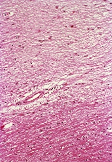
Background imageCentral Nervous System Collection: Multiple sclerosis, light micrograph

Multiple sclerosis, light micrograph
Multiple sclerosis. Light micrograph of a section through the brain showing a large area of demyelinated nerve fibres (light pink, top) due to multiple sclerosis

Background imageCentral Nervous System Collection: Brain glial cells, light micrograph C016 / 0524

Brain glial cells, light micrograph C016 / 0524
Brain glial cells. Light micrograph of a section through brain tissue, showing numerous glial cells (dark). Glial cells, or neuroglia, are non-neural cells of the central nervous system (CNS)

Background imageCentral Nervous System Collection: Brain glial cells, light micrograph C016 / 0523

Brain glial cells, light micrograph C016 / 0523
Brain glial cells. Light micrograph of a section through brain tissue, showing numerous glial cells (dark). Glial cells, or neuroglia, are non-neural cells of the central nervous system (CNS)

Background imageCentral Nervous System Collection: Corpus callosum, DTI MRI scan C017 / 7045

Corpus callosum, DTI MRI scan C017 / 7045
Corpus callosum. Coronal 3D diffusion tensor imaging (DTI) magnetic resonance imaging (MRI) scan of nerve pathways (blue) in and around the brains corpus callosum

Background imageCentral Nervous System Collection: Brain scan, conceptual image C017 / 7393

Brain scan, conceptual image C017 / 7393
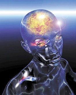
MODEL RELEASED. Brain scan, conceptual composite image

Background imageCentral Nervous System Collection: Human brain, artwork C017 / 7399

Human brain, artwork C017 / 7399
Human brain, computer artwork

Background imageCentral Nervous System Collection: Brain scan, conceptual image C017 / 7396

Brain scan, conceptual image C017 / 7396
MODEL RELEASED. Brain scan, conceptual composite image

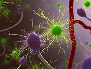
Background imageCentral Nervous System Collection: Nervous system cells, artwork C017 / 3423

Nervous system cells, artwork C017 / 3423
Nervous system cells. Computer artwork showing neurons (nerve cells, purple), astrocytes (green) and a blood vessel (along right)

Background imageCentral Nervous System Collection: Glioblastoma brain tumour, DTI MRI scan C017 / 7052

Glioblastoma brain tumour, DTI MRI scan C017 / 7052
Glioblastoma brain tumour. 3D diffusion tensor imaging (DTI) magnetic resonance imaging (MRI) scan of nerve pathways (coloured) around a glioblastoma (upper right) in the brain

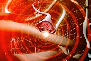
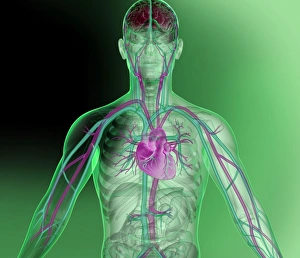
Background imageCentral Nervous System Collection: Heart and brain vascular system, artwork

Heart and brain vascular system, artwork
Heart and brain vascular system. Artwork showing the arteries (pink) and veins (blue) of the torso, arms, neck, head and brain. The brain is at top (red) and the heart at centre

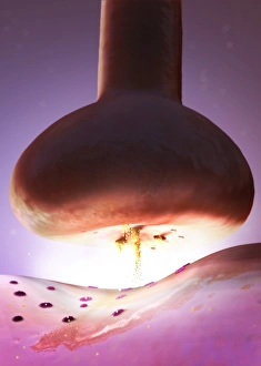
Background imageCentral Nervous System Collection: Nerve synapse, artwork C017 / 3426

Nerve synapse, artwork C017 / 3426
Nerve synapse. Computer artwork of of a junction, or synapse, between two nerve cells (neurons). As the electrical signal reaches the presynaptic end of a neuron it triggers the release of

Background imageCentral Nervous System Collection: Microglial white blood cell, SEM C019 / 0247

Microglial white blood cell, SEM C019 / 0247
Microglial white blood cell, SEM

Background imageCentral Nervous System Collection: Microglial white blood cell, SEM C019 / 0246

Microglial white blood cell, SEM C019 / 0246
Microglia. Coloured scanning electron micrograph (SEM) of an activated microglial white blood cell. This immune system cell is found in the brain and spinal cord

Background imageCentral Nervous System Collection: Microglial white blood cell, SEM C019 / 0245

Microglial white blood cell, SEM C019 / 0245
Microglia. Coloured scanning electron micrograph (SEM) of an activated microglial white blood cell. This immune system cell is found in the brain and spinal cord

Background imageCentral Nervous System Collection: Psychic brain, conceptual image C014 / 1480

Psychic brain, conceptual image C014 / 1480
Psychic brain, conceptual image. Artwork showing the human brain from the side, surrounded by flames, with the pineal gland shown

Background imageCentral Nervous System Collection: Psychic brain, conceptual image C014 / 1478

Psychic brain, conceptual image C014 / 1478
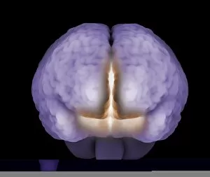
Psychic brain, conceptual image. Artwork of an anterior (frontal) view of the human brain. The cerebrum (the main visible area) is divided into two hemispheres

Background imageCentral Nervous System Collection: Psychic brain, conceptual image C014 / 1479

Psychic brain, conceptual image C014 / 1479
Psychic brain, conceptual image. Artwork of an anterior (frontal) view of the human brain. The cerebrum (the main visible area) is divided into two hemispheres

Background imageCentral Nervous System Collection: Human brain on walking man, artwork

Human brain on walking man, artwork
Human brain on walking man. Artwork showing a brain on a walking man. The brain is the core component of the central nervous system (CNS), with the other part being the spinal cord

Background imageCentral Nervous System Collection: Central and peripheral nerves, artwork

Central and peripheral nerves, artwork
Central and peripheral nerves. Artwork showing the central and peripheral nervous systems (right), with the brain shown on the walking man at left

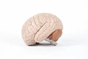
Background imageCentral Nervous System Collection: Human brain, anatomical model

Human brain, anatomical model
Human brain, model. Model (from 2000), showing the outer structure of the human brain

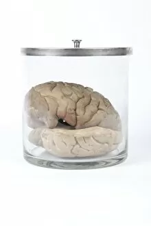
Background imageCentral Nervous System Collection: Human brain, historical anatomical model

Human brain, historical anatomical model
Human brain. 1960s anatomical model of a human brain in a jar

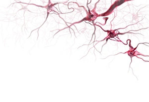
Background imageCentral Nervous System Collection: Brain cells, artwork

Brain cells, artwork
Brain cells. Computer artwork representing neurons (nerve cells) in the brain

Background imageCentral Nervous System Collection: Brain nerve cells, TEM C014 / 0356

Brain nerve cells, TEM C014 / 0356
Brain nerve cells. Transmission electron micrograph (TEM) of a section through brain tissue from the cerebral cortex, showing numerous neurons (nerve cells) surrounded by axons and dendrites

Background imageCentral Nervous System Collection: Brain cell, TEM C014 / 0358

Brain cell, TEM C014 / 0358
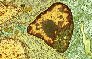
Brain cell. Transmission electron micrograph (TEM) of a section through an oligodendrocyte in human brain tissue. Oligodendrocytes occur in both the white

Background imageCentral Nervous System Collection: Brain cell, TEM C014 / 0359

Brain cell, TEM C014 / 0359
Brain cell. Transmission electron micrograph (TEM) of a section through an oligodendrocyte in human brain tissue. Oligodendrocytes occur in both the white

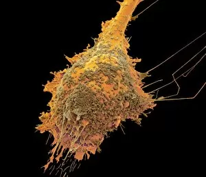
Background imageCentral Nervous System Collection: Dividing brain cancer cells, SEM C014 / 0354

Dividing brain cancer cells, SEM C014 / 0354
Dividing brain cancer cells. Coloured scanning electron micrograph (SEM) of a cancerous astrocyte brain cell that has just undergone cytokinesis (cell division)

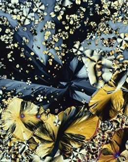
Background imageCentral Nervous System Collection: Melatonin, light micrograph

Melatonin, light micrograph
Melatonin. Light micrograph in polarised light of crystals of the hormone melatonin. Melatonin is secreted by the pineal gland in the brain that controls the bodys biological rhythm

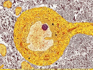
Background imageCentral Nervous System Collection: Purkinje nerve cell, TEM C014 / 0582

Purkinje nerve cell, TEM C014 / 0582
Purkinje nerve cell. Transmission electron micrograph (TEM) of a purkinje nerve cell (bright yellow, centre) from the cerebellum of the brain

Background imageCentral Nervous System Collection: Ionotropic glutamate receptor C015 / 5813

Ionotropic glutamate receptor C015 / 5813
Ionotropic glutamate receptor, molecular model. When glutamate binds to this receptor, it opens up trans-membrane ion channels vital for the functioning of the nervous system

Background imageCentral Nervous System Collection: Ionotropic glutamate receptor C015 / 5812

Ionotropic glutamate receptor C015 / 5812
Ionotropic glutamate receptor, molecular model. When glutamate binds to this receptor, it opens up trans-membrane ion channels vital for the functioning of the nervous system

Background imageCentral Nervous System Collection: Brain, artwork C014 / 6803

Brain, artwork C014 / 6803
Brain, computer artwork. At bottom is the brainstem, which controls automatic functions, sleep and arousal and relays messages from the brain to the spinal cord

Background imageCentral Nervous System Collection: Central nervous system, artwork

Central nervous system, artwork
Central nervous system. Computer artwork showing the brain, cranial nerves and the top portion of the spinal cord. At centre is the pituitary gland (small, spherical), which secretes hormones

Background imageCentral Nervous System Collection: Cyborg brain, artwork C016 / 5792

Cyborg brain, artwork C016 / 5792
Cyborg brain. Artwork of a humanoid with the brain highlighted. The merging of humans and technology is called cybernetics, and the result is known as a cyborg

Background imageCentral Nervous System Collection: Marie Jean Pierre Flourens (1794-1867)

Marie Jean Pierre Flourens (1794-1867)
Celebrated French physiologist, who became Secretary of the Academy of Sciences, accepted the Professorship of Natural History at the College of France

Background imageCentral Nervous System Collection: Synapses, artwork C014 / 0002

Synapses, artwork C014 / 0002
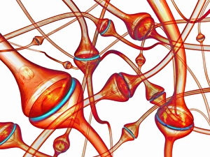
Synapses. Computer artwork of synapses, the junction between nerve cells (orange). Synapses transmit electrical signals from one nerve cell to the next

Background imageCentral Nervous System Collection: Synapses, artwork C014 / 0004

Synapses, artwork C014 / 0004
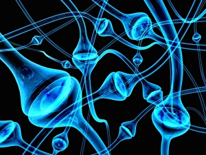
Synapses. Computer artwork of synapses, the junction between nerve cells (blue). Synapses transmit electrical signals from one nerve cell to the next

Background imageCentral Nervous System Collection: Synapses, artwork C014 / 0003

Synapses, artwork C014 / 0003
Synapses. Computer artwork of synapses, the junction between nerve cells (blue). Synapses transmit electrical signals from one nerve cell to the next


All Professionally Made to Order for Quick Shipping

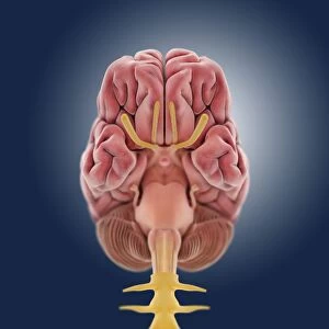
The central nervous system, the intricate network that governs our every thought and movement, is a marvel of complexity. From the delicate cerebellum tissue to the detailed light micrograph capturing its essence, we are reminded of its importance in maintaining balance and coordination. Anatomy comes alive as we explore the human brain from an inferior view. The intricacy of brain fibers is revealed through DTI MRI scans like C017/7099 and C017/7035, showcasing their vital role in transmitting information throughout this extraordinary organ. Artistic renderings bring us closer to understanding the medulla oblongata's significance within the brain. Its portrayal in various artworks allows us to appreciate how it controls essential functions such as breathing and heart rate. As we delve deeper into studying the central nervous system, models of the human brain provide invaluable insights into its structure and organization. Lateral views reveal countless regions responsible for cognition, emotion regulation, sensory perception, and motor control. Microscope slides offer glimpses into nerve cells' intricate architecture—a testament to their ability to transmit electrical signals at lightning speed. Meanwhile, glial stem cell cultures captured under a light microscope remind us of their crucial role in supporting neuronal function. Finally, artistic representations unveil the limbic system's enigmatic nature—an interconnected web responsible for emotions and memory formation. These captivating artworks allow us to visualize this complex network within our brains. Exploring these hints provides a glimpse into the awe-inspiring world of our central nervous system—the very foundation upon which our thoughts, actions, memories reside—reminding us just how remarkable our brains truly are.