Fraud Blocker Skip to main content

Biomolecule Collection (page 6)

"Biomolecules: Unveiling the Intricate World of Life's Building Blocks" Peering through the lens of scientific discovery

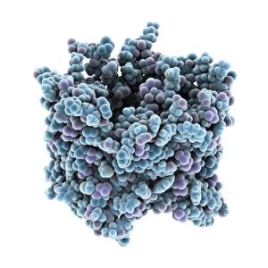
Background imageBiomolecule Collection: MUC7 molecule

MUC7 molecule. Computer model showing the structure of a molecule of the protein MUC7 (mucin 7). Mucins key characteristic is their ability to form gels

Background imageBiomolecule Collection: Ebola virus transcription factor fragment

Ebola virus transcription factor fragment. Molecular model of the C-terminal domain (CTD) of Ebola virus transcription factor VP30

Background imageBiomolecule Collection: Bacterial twitching motility protein

Bacterial twitching motility protein
pilt, , protein, biomolecule, macromolecule, bacterial twitching motility, enzyme, aquifex aeolicus, bacterium, biochemistry, biology, molecular biology, proteomics, bacteriology, microbiology

Background imageBiomolecule Collection: RuvBL1 helicase enzyme

RuvBL1 helicase enzyme, molecular model. Helicases are enzymes that carry out several roles, primarily separating the two strands of the DNA (deoxyribonucleic acid) double helix

Background imageBiomolecule Collection: Metal-binding protein bound to DNA

Metal-binding protein bound to DNA. Molecular model of the bacterial metal-binding protein NikR (bottom) bound to a strand of DNA (top, helical, deoxyribonucleic acid)

Background imageBiomolecule Collection: Muscle contraction proteins

Muscle contraction proteins. Molecular model of muscle protein motor cross-bridges during contraction in muscle. The cross-bridge is seen from the side, with contraction taking place horizontally

Background imageBiomolecule Collection: MUC5B molecule

MUC5B molecule. Computer model showing the structure of a molecule of the protein MUC5B (mucin 5 subtype B). Mucins key characteristic is their ability to form gels

Background imageBiomolecule Collection: Myoglobin protein, molecular model C016 / 6575

Myoglobin protein, molecular model C016 / 6575
Myoglobin protein. Molecular model showing the structure of the myoglobin protein. Myoglobin is a protein found in muscle tissue

Background imageBiomolecule Collection: Alpha-beta T-cell receptor

Alpha-beta T-cell receptor, molecular model. The T cell receptor (TCR) is a protein complex found on the surface of a type of white blood cell called T lymphocytes (or T cells)

Background imageBiomolecule Collection: Carbamoylsarcosine amidase enzyme

Carbamoylsarcosine amidase enzyme, molecular model. This enzyme catalyses the chemical reaction that converts the chemical N-carbamoylsarcosine in the presence of water to sarcosine (N-methylglycine)

Background imageBiomolecule Collection: Genomic HIV-RNA duplex

Genomic HIV-RNA duplex, molecular model. This structure shows the dimerization initiation site of genomic HIV-1 with RNA (ribonucleic acid)

Background imageBiomolecule Collection: Transducin protein beta-gamma complex

Transducin protein beta-gamma complex. Molecular model of the beta-gamma dimer of the heterotrimeric G protein transducin

Background imageBiomolecule Collection: Nerve growth factor bound to receptor

Nerve growth factor bound to receptor, molecular model. Nerve growth factor (NGF) complexed with the TrkA receptor. NGF is a neurotrophin that acts on the development and function of nerves

Background imageBiomolecule Collection: Chromosome segregation protein

Chromosome segregation protein, molecular model. This proteins function is to aid the process of chromosome segregation during cell division and replication

Background imageBiomolecule Collection: Nerve growth factor protein complex

Nerve growth factor protein complex, molecular model. This complex consists of nerve growth factor (NGF) in complex with four binding proteins

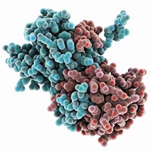
Background imageBiomolecule Collection: Thymidylic acid-ribonuclease A complex

Thymidylic acid-ribonuclease A complex. Molecular model of a thymidylic acid tetramer (blue) in complex with ribonuclease A (red)

Background imageBiomolecule Collection: DNA bundle on silicon nanopillars, SEM

DNA bundle on silicon nanopillars, SEM
DNA bundle on silicon nanopillars. Scanning electron micrograph (SEM) of a DNA (deoxyribonucleic acid) bundle and silicon nanopillars used to obtain the first high-contrast direct images of DNA

Background imageBiomolecule Collection: Exosome complex, molecular model

Exosome complex, molecular model. This multi-protein complex functions to break up strands of RNA (ribonucleic acid, pink) during biochemical processes

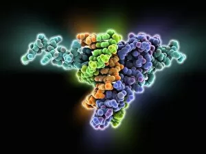
Background imageBiomolecule Collection: MyoD muscle protein-DNA complex

MyoD muscle protein-DNA complex. Molecular model of the MyoD muscle protein (green and blue) bound to a strand of DNA (deoxyribonucleic acid, orange and green)

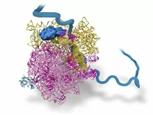
Background imageBiomolecule Collection: Bacterial ribosome and protein synthesis

Bacterial ribosome and protein synthesis. Molecular model showing a bacterial ribosome reading an mRNA (messenger ribonucleic acid) strand (blue) and synthesising a protein

Background imageBiomolecule Collection: Canine parvovirus capsid C015 / 8460

Canine parvovirus capsid C015 / 8460
Canine parvovirus capsid, molecular model. Canine parvovirus type 2 (CPV2) was first recognized in 1978 and spread worldwide in less than two years

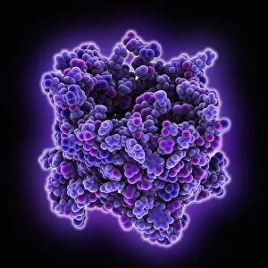
Background imageBiomolecule Collection: VDAC-1 ion channel protein C015 / 8250

VDAC-1 ion channel protein C015 / 8250
VDAC-1 ion channel protein, molecular model. This is the human voltage-dependent anion-selective channel protein 1 (VDAC-1)

Background imageBiomolecule Collection: Streptavidin bacterial protein C015 / 8458

Streptavidin bacterial protein C015 / 8458
Streptavidin bacterial protein, molecular model. Streptavidin is a protein obtained from the bacterium Streptomyces avidinii

Background imageBiomolecule Collection: Calcium ATPase ion pump C015 / 7143

Calcium ATPase ion pump C015 / 7143
Calcium ATPase ion pump, molecular model. This enzyme is found in muscle cell membranes, where it pumps calcium in and out of muscle cells and controls muscle contractions

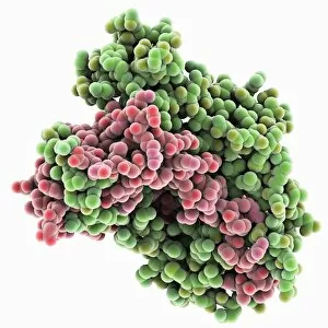
Background imageBiomolecule Collection: DNA repair protein AlkB with DNA C016 / 0547

DNA repair protein AlkB with DNA C016 / 0547
DNA repair protein AlkB with DNA. Molecular model of the DNA (deoxyribonucleic acid) repair protein AlkB (blue) bound to a strand of double-stranded DNA (ds-DNA, pink and yellow)

Background imageBiomolecule Collection: Sodium-potassium ion pump proteins C015 / 9993

Sodium-potassium ion pump proteins C015 / 9993
Sodium-potassium ion pump proteins, molecular model. Sodium-potassium ATPase (adenosine triphosphatase) is an ATP-powered ion pump found in all animal cells

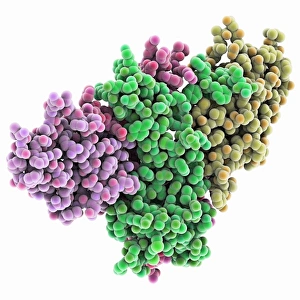
Background imageBiomolecule Collection: Thrombin complexed with fibrinogen C015 / 7148

Thrombin complexed with fibrinogen C015 / 7148
Thrombin complexed with fibrinogen, molecular model. The thrombin molecules (left and right, purple and green) are bound to the central part of the fibrinogen molecule (centre, multiple colours)

Background imageBiomolecule Collection: 3-hydroxyacyl-CoA dehydrogenase C015 / 9940

3-hydroxyacyl-CoA dehydrogenase C015 / 9940
3-hydroxyacyl-CoA dehydrogenase, molecular model. This enzyme is found in human heart tissue, and catalyzes a reaction that is part of the beta-oxidation pathway

Background imageBiomolecule Collection: Streptavidin bacterial protein C015 / 8457

Streptavidin bacterial protein C015 / 8457
Streptavidin bacterial protein, molecular model. Streptavidin is a protein obtained from the bacterium Streptomyces avidinii

Background imageBiomolecule Collection: Sodium-potassium ion pump proteins C015 / 9997

Sodium-potassium ion pump proteins C015 / 9997
Sodium-potassium ion pump proteins, molecular model. Sodium-potassium ATPase (adenosine triphosphatase) is an ATP-powered ion pump found in all animal cells

Background imageBiomolecule Collection: Retinal-producing oxygenase enzyme C015 / 7150

Retinal-producing oxygenase enzyme C015 / 7150
Retinal-producing oxygenase enzyme, molecular model. This enzyme, an oxygenase, plays a role in the production and metabolism of retinal and other apocarotenoids

Background imageBiomolecule Collection: Central glycolytic gene regulator protein C016 / 0549

Central glycolytic gene regulator protein C016 / 0549
Central glycolytic gene regulator (CGGR) protein, molecular model. This protein binds to DNA (deoxyribonucleic acid) in the absence of glucose, blocking the transcription of certain genes

Background imageBiomolecule Collection: Haemoglobin, molecular model C015 / 9391

Haemoglobin, molecular model C015 / 9391
Haemoglobin, molecular model. This is deoxyhaemoglobin, the molecule in its non-oxygen bound state. Haemoglobin transports oxygen around the body in red blood cells

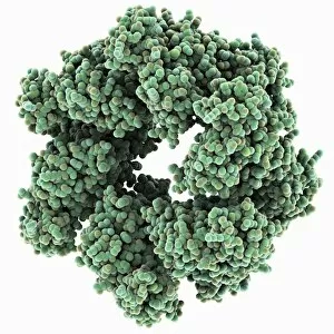
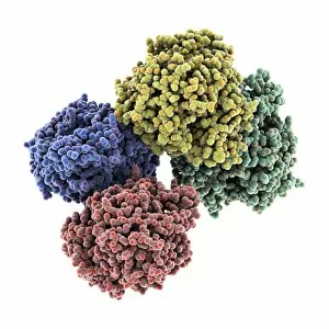
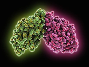
Background imageBiomolecule Collection: Hemolysin-coregulated protein

Hemolysin-coregulated protein, molecular model. This protein is from the bacterium Pseudomonas aeruginosa. It is a hexameric ring structure, named hcp1

Background imageBiomolecule Collection: Canine parvovirus capsid C015 / 8459

Canine parvovirus capsid C015 / 8459
Canine parvovirus capsid, molecular model. Canine parvovirus type 2 (CPV2) was first recognized in 1978 and spread worldwide in less than two years

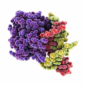
Background imageBiomolecule Collection: DNA repair protein AlkB with DNA C016 / 0546

DNA repair protein AlkB with DNA C016 / 0546
DNA repair protein AlkB with DNA. Molecular model of the DNA (deoxyribonucleic acid) repair protein AlkB (purple) bound to a strand of double-stranded DNA (ds-DNA, red and green)

Background imageBiomolecule Collection: Interferon-DNA transcription complex C015 / 8251

Interferon-DNA transcription complex C015 / 8251
Interferon-DNA transcription complex, molecular model. Bound to the DNA (deoxyribonucleic acid, green and yellow) is transcription factor p65, interferon regulatory factor 7

Background imageBiomolecule Collection: Interferon-DNA transcription complex C015 / 8252

Interferon-DNA transcription complex C015 / 8252
Interferon-DNA transcription complex, molecular model. Bound to the DNA (deoxyribonucleic acid, pink and white) is transcription factor p65, interferon regulatory factor 7, interferon fusion protein

Background imageBiomolecule Collection: Vitamin B12 import proteins C015 / 9942

Vitamin B12 import proteins C015 / 9942
Vitamin B12 import proteins, molecular model. This complex is the import proteins btuC, btuD, and btuF. The first two together form BtuCD

Background imageBiomolecule Collection: VDAC-1 ion channel protein C015 / 8249

VDAC-1 ion channel protein C015 / 8249
VDAC-1 ion channel protein, molecular model. This is the human voltage-dependent anion-selective channel protein 1 (VDAC-1)

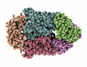
Background imageBiomolecule Collection: Thrombin complexed with fibrinogen C015 / 7149

Thrombin complexed with fibrinogen C015 / 7149
Thrombin complexed with fibrinogen, molecular model. The thrombin molecules (left and right, brown and pink) are bound to the central part of the fibrinogen molecule (centre, multiple colours)

Background imageBiomolecule Collection: Simian virus (SV40) large T antigen C015 / 7069

Simian virus (SV40) large T antigen C015 / 7069
Simian virus (SV40) large T antigen, molecular model. This antigen is from the simian vacuolating virus 40 (SV40). Large T antigens play a role in regulating the viral life cycle of

Background imageBiomolecule Collection: Haemoglobin, molecular model C015 / 9392

Haemoglobin, molecular model C015 / 9392
Haemoglobin, molecular model. This is deoxyhaemoglobin, the molecule in its non-oxygen bound state. Haemoglobin transports oxygen around the body in red blood cells

Background imageBiomolecule Collection: 3-hydroxyacyl-CoA dehydrogenase C015 / 9941

3-hydroxyacyl-CoA dehydrogenase C015 / 9941
3-hydroxyacyl-CoA dehydrogenase, molecular model. This enzyme is found in human heart tissue, and catalyzes a reaction that is part of the beta-oxidation pathway

Background imageBiomolecule Collection: Vitamin B12 import proteins C015 / 9943

Vitamin B12 import proteins C015 / 9943
Vitamin B12 import proteins, molecular model. This complex is the import proteins btuC, btuD, and btuF. The first two together form BtuCD

Background imageBiomolecule Collection: Calcium ATPase ion pump C015 / 7142

Calcium ATPase ion pump C015 / 7142
Calcium ATPase ion pump, molecular model. This enzyme is found in muscle cell membranes, where it pumps calcium in and out of muscle cells and controls muscle contractions

Background imageBiomolecule Collection: Retinal-producing oxygenase enzyme C015 / 7151

Retinal-producing oxygenase enzyme C015 / 7151
Retinal-producing oxygenase enzyme, molecular model. This enzyme, an oxygenase, plays a role in the production and metabolism of retinal and other apocarotenoids

Background imageBiomolecule Collection: Adeno-associated virus, molecular model C018 / 0449

Adeno-associated virus, molecular model C018 / 0449
Adeno-associated virus (aV), molecular model. This image shows the core protein of the non-enveloped virus that surrounds the genetic material


All Professionally Made to Order for Quick Shipping

"Biomolecules: Unveiling the Intricate World of Life's Building Blocks" Peering through the lens of scientific discovery, we witness a microscopic view of the human respiratory syncytial virus, unraveling its intricate structure and shedding light on its mechanisms. Zinc fingers delicately embrace a DNA strand, forming an elegant dance between proteins and genetic material. This interaction holds the key to unlocking the mysteries encoded within our genes. The SARS coronavirus protein stands as a formidable foe in our battle against infectious diseases. Understanding its structure is crucial for developing effective treatments and vaccines. Illustrated with precision, a protein takes center stage, showcasing nature's artistic design. Its complex folds and twists hold immense power in shaping life's diverse forms and functions. TFAM transcription factor gracefully binds to DNA C015/7059, orchestrating gene expression like a conductor leading an orchestra. This molecular symphony plays a vital role in maintaining cellular harmony. Stylized rabies virus particles remind us of nature's ability to create both beauty and danger simultaneously. The intricacies hidden within these tiny entities have captivated scientists for centuries. A glimpse into the microscopic world reveals yellow fever virus particles resembling vibrant orbs dancing amidst darkness—a reminder that even invisible threats can wield significant impact on human health. Conceptual imagery portrays the enigmatic rabies virus—an embodiment of fear lurking in shadows yet inspiring relentless pursuit towards understanding this deadly pathogen. MyoD muscle protein-DNA complex showcases how molecules choreograph muscle development—binding together like partners engaged in an exquisite ballet that shapes our physical strength. MscL ion channel protein structure presents itself as nature's gateway—allowing ions to traverse cell membranes with precise control over vital processes essential for life itself. Adenovirus hexon protein emerges as an architectural masterpiece—a structural cornerstone dictating viral entry into host cells while captivating researchers seeking innovative antiviral strategies.