Fraud Blocker Skip to main content

Biomolecule Collection (page 4)

"Biomolecules: Unveiling the Intricate World of Life's Building Blocks" Peering through the lens of scientific discovery

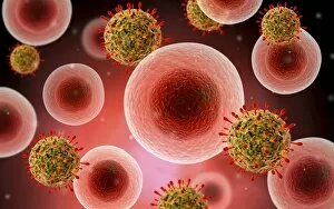
Background imageBiomolecule Collection: Microscopic view of cell and virus

Microscopic view of cell and virus

Background imageBiomolecule Collection: Microscopic view of blood cells

Microscopic view of blood cells

Background imageBiomolecule Collection: Conceptual image of a blood vessel

Conceptual image of a blood vessel

Background imageBiomolecule Collection: Conceptual image of red blood cells

Conceptual image of red blood cells

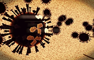
Background imageBiomolecule Collection: A black swarm of H5N1 avian flu viruses are attacked by antibodies

A black swarm of H5N1 avian flu viruses are attacked by antibodies (the three legged elements), which mark the virus for destruction

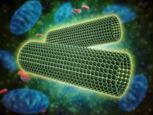
Background imageBiomolecule Collection: Microscopic view of centrioles within a human cell

Microscopic view of centrioles within a human cell
Microscopic view of barrel shaped structure of centrioles within a human cell. A centriole is a cylinder shaped cell structure found in most eukaryotic cells

Background imageBiomolecule Collection: Conceptual image of bacteria

Conceptual image of bacteria

Background imageBiomolecule Collection: Conceptual image of common virus

Conceptual image of common virus

Background imageBiomolecule Collection: Components of blood vessel interact with intracellular fluid

Components of blood vessel interact with intracellular fluid; white blood cells, red blood cells, oxygen, platelets and hormones

Background imageBiomolecule Collection: Microscopic view of Yersinia

Microscopic view of Yersinia. Yersinia is a gram-negative, rod-shaped bacteria in the family Enterobacteriaceae

Background imageBiomolecule Collection: Microscopic view of a microbe

Microscopic view of a microbe. Microbes are single-cell organisms so tiny that millions can fit into the eye of a needle

Background imageBiomolecule Collection: Conceptual image of Basophils

Conceptual image of Basophils. Basophils are part of your immune system that normally protects your body from infection, but can also be partly responsible for your asthma symptoms

Background imageBiomolecule Collection: Microscopic view of bacteria

Microscopic view of bacteria

Background imageBiomolecule Collection: Malria spores in the human blood stream

Malria spores in the human blood stream
The malaria spores move from the mosquito to the human blood stream. the spores divide many times and produce large numbers of spores

Background imageBiomolecule Collection: Conceptual image of lyssavirus

Conceptual image of lyssavirus. Lyssavirus is a genus of viruses belonging to the family Rhabdoviridae. This group of RNA viruses includes the rabies virus traditionally associated with the disease

Background imageBiomolecule Collection: Conceptual image of a ubiquitous virus

Conceptual image of a ubiquitous virus. A ubiquitous virus is contagious in early childhood through the respiratory tract

Background imageBiomolecule Collection: Microscopic view of Henipavirus

Microscopic view of Henipavirus. Henipavirus is a established group of paramyxoviruses comprising the Hendra virus, Nipah virus and Cedar virus

Background imageBiomolecule Collection: Microscopic view of red blood cells flowing inside lungs

Microscopic view of red blood cells flowing inside lungs

Background imageBiomolecule Collection: Artery cross section with red blood cell flow

Artery cross section with red blood cell flow

Background imageBiomolecule Collection: Microscopic view of HIV virus

Microscopic view of HIV virus

Background imageBiomolecule Collection: Red blood cells with white blood cells

Red blood cells with white blood cells

Background imageBiomolecule Collection: Conceptual image of common bacteria

Conceptual image of common bacteria

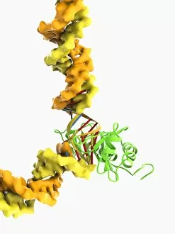
Background imageBiomolecule Collection: TATA box-binding protein complex C017 / 7082

TATA box-binding protein complex C017 / 7082
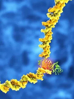
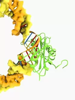
TATA box-binding protein complex. Molecular model showing a TATA box-binding protein (TBP, green) complexed with a strand of DNA (deoxyribonucleic acid, yellow) and transcription factor IIB

Background imageBiomolecule Collection: TATA box-binding protein complex C017 / 7088

TATA box-binding protein complex C017 / 7088
TATA box-binding protein complex. Molecular model showing a TATA box-binding protein (TBP, green) complexed with a strand of DNA (deoxyribonucleic acid, yellow) and transcription factor IIB

Background imageBiomolecule Collection: TATA box-binding protein complex C017 / 7084

TATA box-binding protein complex C017 / 7084
TATA box-binding protein complex. Molecular model showing a TATA box-binding protein (TBP, green) complexed with a strand of DNA (deoxyribonucleic acid, yellow) and transcription factor IIB

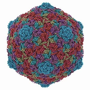
Background imageBiomolecule Collection: HK97 bacteriophage capsid

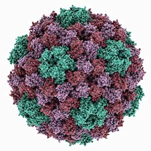
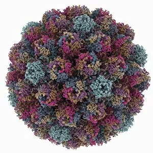
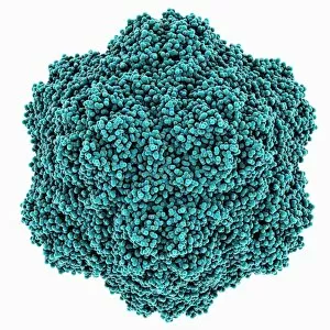
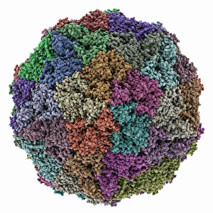
HK97 bacteriophage capsid, molecular model. Bacteriophages are viruses that infect bacteria, in this case enterobacteria such as E. coli (Escherichia coli), with the phage head shown here

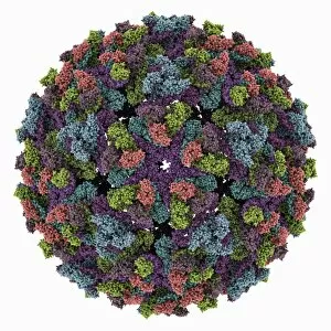
Background imageBiomolecule Collection: Chikungunya virus capsid

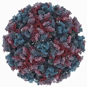
Chikungunya virus capsid, molecular model. This virus, transmitted by mosquitoes in tropical Africa and Asia, causes fever and joint pain in humans, similar to dengue fever

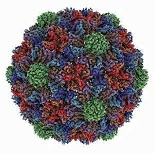
Background imageBiomolecule Collection: HK97 bacteriophage procapsid

HK97 bacteriophage procapsid. Molecular model showing the structure of the prohead-I procapsid of the HK97 bacteriophage. Bacteriophages are viruses that infect bacteria

Background imageBiomolecule Collection: Turnip yellow mosaic virus capsid

Turnip yellow mosaic virus capsid, molecular model. This virus infects a wide variety of plants, including crops such as turnips and cabbages, causing yellow patches on the leaves

Background imageBiomolecule Collection: Sindbis virus capsid, molecular model

Sindbis virus capsid, molecular model. This virus, transmitted by mosquitoes, causes sindbis fever in humans. In viruses, the capsid is the protein shell that encloses the genetic material

Background imageBiomolecule Collection: Murine polyomavirus capsid

Murine polyomavirus capsid, molecular model. This virus, one of a range named for their potential to cause multiple tumours, infects mice

Background imageBiomolecule Collection: Brome mosaic virus capsid

Brome mosaic virus capsid, molecular model. This plant virus infects grasses, especially brome grasses, and also barley. It causes mosaic patches of discolouration

Background imageBiomolecule Collection: Cowpea chlorotic mottle virus capsid

Cowpea chlorotic mottle virus capsid, molecular model. This virus (CCMV) infects the cowpea plant (Vigna unguiculata), causing yellow spots of discolouration

Background imageBiomolecule Collection: Potassium ion channel protein structure

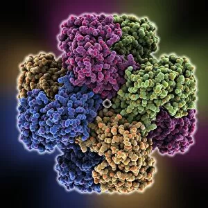
Potassium ion channel protein structure. Molecular model of a KcsA potassium ion (K+) channel from Streptomyces lividans bacteria

Background imageBiomolecule Collection: Streptavidin-biotin molecular complex

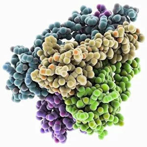
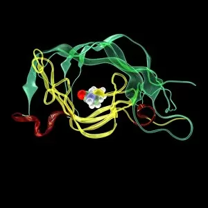
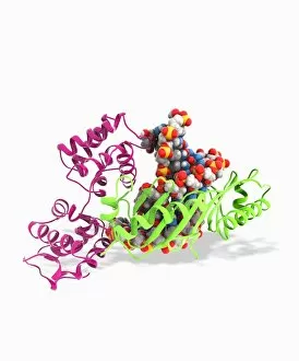
Streptavidin-biotin molecular complex. Molecular model of a single-strand binding complex of streptavidin (ribbons) and biotin (space-filled model, centre). Biotin is also known as vitamin B7

Background imageBiomolecule Collection: Potassium ion channel beta subunit

Potassium ion channel beta subunit. Molecular model showing the structure a beta subunit of a voltage-dependent potassium (K+) channel

Background imageBiomolecule Collection: KCNQ ion channel protein structure

KCNQ ion channel protein structure. Molecular model showing the protein structure of an ion channel domain. Ion channels are membrane-spanning proteins that form a pathway for the movement of

Background imageBiomolecule Collection: Potassium ion channel cavity structure

Potassium ion channel cavity structure. Molecular model showing the structure of a cavity formed by potassium ion channel proteins

Background imageBiomolecule Collection: Avian polyomavirus capsid

Avian polyomavirus capsid, molecular model. This virus, one of a range named for their potential to cause multiple tumours, infects birds. Discovered in budgerigars in 1981, it is often fatal

Background imageBiomolecule Collection: Cytoplasmic polyhedrosis virus capsid

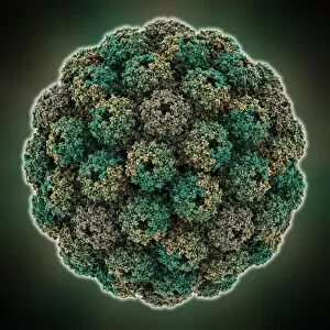
Cytoplasmic polyhedrosis virus capsid, molecular model. Part of the Cypovirus genus and invariably fatal, this insect virus is transmitted by contamination of leaves eaten (examples include silkworms)

Background imageBiomolecule Collection: TATA box-binding protein complex C017 / 7090

TATA box-binding protein complex C017 / 7090
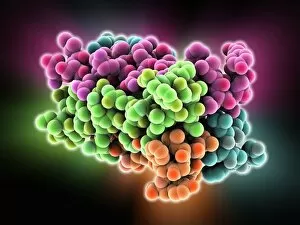
TATA box-binding protein complex. Molecular model showing a TATA box-binding protein (TBP, green) complexed with a strand of DNA (deoxyribonucleic acid, spheres) and transcription factor IIB

Background imageBiomolecule Collection: Theilers encephalomyelitis virus capsid

Theilers encephalomyelitis virus capsid, molecular model. This virus, which causes brain and spinal cord inflammation in mice, is used in research

Background imageBiomolecule Collection: TATA box-binding protein complex C017 / 7085

TATA box-binding protein complex C017 / 7085
TATA box-binding protein complex. Molecular model showing a TATA box-binding protein (TBP, green) complexed with a strand of DNA (deoxyribonucleic acid, yellow) and transcription factor IIB

Background imageBiomolecule Collection: Tobacco necrosis virus capsid

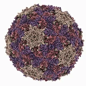
Tobacco necrosis virus capsid, molecular model. This plant virus infects a wide rage of plants, including the tobacco plant for which it is named. The virus causes tissue death (necrosis)

Background imageBiomolecule Collection: TATA box-binding protein complex C017 / 7083

TATA box-binding protein complex C017 / 7083
TATA box-binding protein complex. Molecular model showing a TATA box-binding protein (TBP, green) complexed with a strand of DNA (deoxyribonucleic acid, yellow) and transcription factor IIB

Background imageBiomolecule Collection: Grapevine fanleaf virus capsid

Grapevine fanleaf virus capsid, molecular model. This plant virus is named for its infection of grape vines. It is transmitted by the nematode worm Xiphinema index

Background imageBiomolecule Collection: VEE equine encephalitis virus capsid

VEE equine encephalitis virus capsid
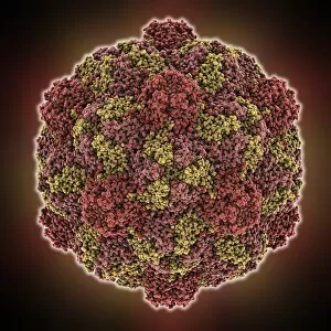
Venezuelan equine encephalitis virus capsid, molecular model. This mosquito-borne virus can kill horses and other equine species, causing brain and spinal cord inflammation

Background imageBiomolecule Collection: Murine minute virus capsid

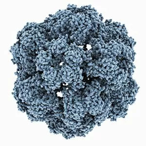
Murine minute virus capsid, molecular model. This parvovirus infects mice, its only known natural host. It is highly infectious, transmitted through the nose and mouth


All Professionally Made to Order for Quick Shipping

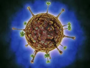
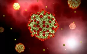
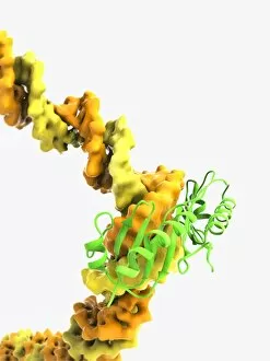
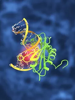
"Biomolecules: Unveiling the Intricate World of Life's Building Blocks" Peering through the lens of scientific discovery, we witness a microscopic view of the human respiratory syncytial virus, unraveling its intricate structure and shedding light on its mechanisms. Zinc fingers delicately embrace a DNA strand, forming an elegant dance between proteins and genetic material. This interaction holds the key to unlocking the mysteries encoded within our genes. The SARS coronavirus protein stands as a formidable foe in our battle against infectious diseases. Understanding its structure is crucial for developing effective treatments and vaccines. Illustrated with precision, a protein takes center stage, showcasing nature's artistic design. Its complex folds and twists hold immense power in shaping life's diverse forms and functions. TFAM transcription factor gracefully binds to DNA C015/7059, orchestrating gene expression like a conductor leading an orchestra. This molecular symphony plays a vital role in maintaining cellular harmony. Stylized rabies virus particles remind us of nature's ability to create both beauty and danger simultaneously. The intricacies hidden within these tiny entities have captivated scientists for centuries. A glimpse into the microscopic world reveals yellow fever virus particles resembling vibrant orbs dancing amidst darkness—a reminder that even invisible threats can wield significant impact on human health. Conceptual imagery portrays the enigmatic rabies virus—an embodiment of fear lurking in shadows yet inspiring relentless pursuit towards understanding this deadly pathogen. MyoD muscle protein-DNA complex showcases how molecules choreograph muscle development—binding together like partners engaged in an exquisite ballet that shapes our physical strength. MscL ion channel protein structure presents itself as nature's gateway—allowing ions to traverse cell membranes with precise control over vital processes essential for life itself. Adenovirus hexon protein emerges as an architectural masterpiece—a structural cornerstone dictating viral entry into host cells while captivating researchers seeking innovative antiviral strategies.