Fraud Blocker Skip to main content

Biomolecule Collection (page 3)

"Biomolecules: Unveiling the Intricate World of Life's Building Blocks" Peering through the lens of scientific discovery

Background imageBiomolecule Collection: Group of H5N1 virus with glassy view

Group of H5N1 virus with glassy view

Background imageBiomolecule Collection: Microscopic view of fat plaque inside the artery

Microscopic view of fat plaque inside the artery

Background imageBiomolecule Collection: Microscopic view of blood flow inside an artery with stent deployment

Microscopic view of blood flow inside an artery with stent deployment

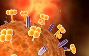
Background imageBiomolecule Collection: Swine influenza virus

Swine influenza virus

Background imageBiomolecule Collection: Microscopic view of a human antibody

Microscopic view of a human antibody. Human antibodies are the Y-shaped proteins used by the immune system to defend against foreign objects like bacteria and viruses

Background imageBiomolecule Collection: Microscopic view of immunoglobulin G antibodies

Microscopic view of immunoglobulin G antibodies

Background imageBiomolecule Collection: The interior of an eukaryotic cell

The interior of an eukaryotic cell. This is the most common generic type of cell and its present in all mammals. In the center is the nucleus (the perforated sphere)

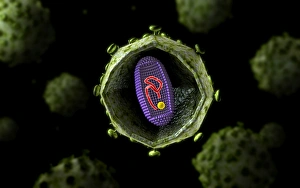
Background imageBiomolecule Collection: Microscopic view of HIV virus, cross section

Microscopic view of HIV virus, cross section

Background imageBiomolecule Collection: Insertion of stent into atherosclerotic artery

Insertion of stent into atherosclerotic artery

Background imageBiomolecule Collection: Artery cross-section with blood flow, fat plaque and stent deployment

Artery cross-section with blood flow, fat plaque and stent deployment
Microscopic view of an artery cross-section with blood flow, fat plaque and stent deployment

Background imageBiomolecule Collection: Conceptual image of Encephalitis

Conceptual image of Encephalitis. Encephalitis can cause flu-like symptoms, such as a fever or severe headaches. It can also cause confused thinking, seizures, or problems with senses

Background imageBiomolecule Collection: Microscopic view of respiratory syncytial virus

Microscopic view of respiratory syncytial virus (RSV). RSV is a common virus that leads to mild, cold-like symptoms in adults and children

Background imageBiomolecule Collection: Conceptual image of red blood cells with platelets

Conceptual image of red blood cells with platelets

Background imageBiomolecule Collection: Microscopic view of HIV virus inside the lungs

Microscopic view of HIV virus inside the lungs

Background imageBiomolecule Collection: Microscopic view of Listeria monocytogenes

Microscopic view of Listeria monocytogenes. Listeria monocytogenes is the causative agent of the bacterial infection called listeriosis

Background imageBiomolecule Collection: Microscopic view of plasma cell inside blood vessel

Microscopic view of plasma cell inside blood vessel

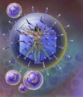
Background imageBiomolecule Collection: Vitruvian Man inside virus particle

Vitruvian Man inside virus particle

Background imageBiomolecule Collection: Inside view of the artery with red blood cells

Inside view of the artery with red blood cells

Background imageBiomolecule Collection: Clathrin Mediated Endocytosis

Clathrin Mediated Endocytosis

Background imageBiomolecule Collection: Microscopic cross section view of the small intestine

Microscopic cross section view of the small intestine. TheAsmall intestineAis a long, highly convoluted tube in the digestive system that absorbs about 90% of the nutrients from the food we eat

Background imageBiomolecule Collection: Conceptual image of red blood cells with fibrin

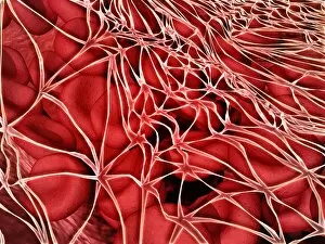
Conceptual image of red blood cells with fibrin

Background imageBiomolecule Collection: Conceptual image of sickle cell anemia

Conceptual image of sickle cell anemia. Sickle cell anemia is a disease in which your body produces abnormally shaped red blood cells. The cells are shaped like a crescent or sickle

Background imageBiomolecule Collection: Microscopic view of bacteriophage

Microscopic view of bacteriophage

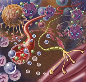
Background imageBiomolecule Collection: Nerve with myelin sheath, seen in lower right, connects with muscle

Nerve with myelin sheath, seen in lower right, connects with muscle. Blood vessel and immune cells are seen in the center and upper right of image

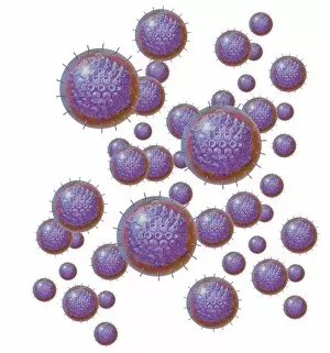
Background imageBiomolecule Collection: Grouping of virus particles

Grouping of virus particles

Background imageBiomolecule Collection: Microscopic view of Rickettsia

Microscopic view of Rickettsia. Rickettsia is a gram-negative bacteria carried by parasites such as ticks, fleas and lice

Background imageBiomolecule Collection: Microscopic view of corncob formation in dental plaque

Microscopic view of corncob formation in dental plaque, believed to be limited to strains of Corynebacterium matruchotii and Streptococcus sanguis

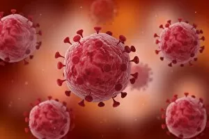
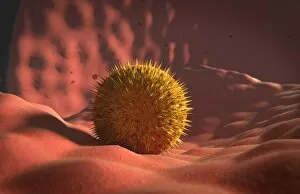
Background imageBiomolecule Collection: Microscopic view of virus

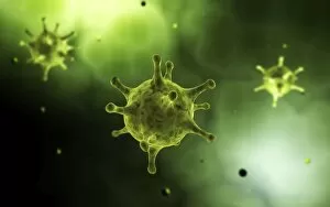
Microscopic view of virus

Background imageBiomolecule Collection: Conceptual image of influenza causing flu virus

Conceptual image of influenza causing flu virus

Background imageBiomolecule Collection: Microscopic view of human anitbodies with red blood cells

Microscopic view of human anitbodies with red blood cells. Human antibodies are the Y-shaped proteins used by the immune system to defend against foreign objects like bacteria and viruses

Background imageBiomolecule Collection: Virus landing on cell membrane

Virus landing on cell membrane

Background imageBiomolecule Collection: Microscopic view of sicke cells causing anemia disease

Microscopic view of sicke cells causing anemia disease

Background imageBiomolecule Collection: Microscopic view of rotavirus

Microscopic view of rotavirus. Rotavirus is the most common cause of severe diarrhea among infants and young children. It is a genus of double-stranded RNA virus in the family Reoviridae

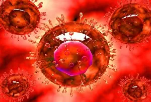
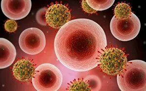
Background imageBiomolecule Collection: Microscopic view of cell and virus

Microscopic view of cell and virus

Background imageBiomolecule Collection: Microscopic view of blood cells

Microscopic view of blood cells

Background imageBiomolecule Collection: Conceptual image of a blood vessel

Conceptual image of a blood vessel

Background imageBiomolecule Collection: Conceptual image of red blood cells

Conceptual image of red blood cells

Background imageBiomolecule Collection: A black swarm of H5N1 avian flu viruses are attacked by antibodies

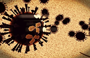
A black swarm of H5N1 avian flu viruses are attacked by antibodies (the three legged elements), which mark the virus for destruction

Background imageBiomolecule Collection: Microscopic view of centrioles within a human cell

Microscopic view of centrioles within a human cell
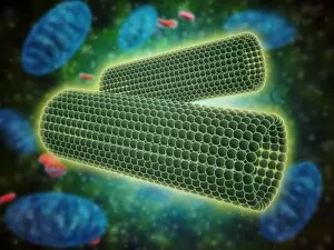
Microscopic view of barrel shaped structure of centrioles within a human cell. A centriole is a cylinder shaped cell structure found in most eukaryotic cells

Background imageBiomolecule Collection: Conceptual image of bacteria

Conceptual image of bacteria

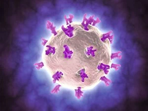
Background imageBiomolecule Collection: Conceptual image of common virus

Conceptual image of common virus

Background imageBiomolecule Collection: Components of blood vessel interact with intracellular fluid

Components of blood vessel interact with intracellular fluid; white blood cells, red blood cells, oxygen, platelets and hormones

Background imageBiomolecule Collection: Microscopic view of Yersinia

Microscopic view of Yersinia. Yersinia is a gram-negative, rod-shaped bacteria in the family Enterobacteriaceae

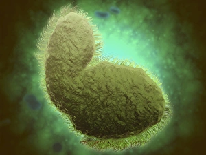
Background imageBiomolecule Collection: Microscopic view of a microbe

Microscopic view of a microbe. Microbes are single-cell organisms so tiny that millions can fit into the eye of a needle

Background imageBiomolecule Collection: Conceptual image of Basophils

Conceptual image of Basophils. Basophils are part of your immune system that normally protects your body from infection, but can also be partly responsible for your asthma symptoms

Background imageBiomolecule Collection: Microscopic view of bacteria

Microscopic view of bacteria

Background imageBiomolecule Collection: Malria spores in the human blood stream

Malria spores in the human blood stream
The malaria spores move from the mosquito to the human blood stream. the spores divide many times and produce large numbers of spores

Background imageBiomolecule Collection: Conceptual image of lyssavirus

Conceptual image of lyssavirus. Lyssavirus is a genus of viruses belonging to the family Rhabdoviridae. This group of RNA viruses includes the rabies virus traditionally associated with the disease


All Professionally Made to Order for Quick Shipping

"Biomolecules: Unveiling the Intricate World of Life's Building Blocks" Peering through the lens of scientific discovery, we witness a microscopic view of the human respiratory syncytial virus, unraveling its intricate structure and shedding light on its mechanisms. Zinc fingers delicately embrace a DNA strand, forming an elegant dance between proteins and genetic material. This interaction holds the key to unlocking the mysteries encoded within our genes. The SARS coronavirus protein stands as a formidable foe in our battle against infectious diseases. Understanding its structure is crucial for developing effective treatments and vaccines. Illustrated with precision, a protein takes center stage, showcasing nature's artistic design. Its complex folds and twists hold immense power in shaping life's diverse forms and functions. TFAM transcription factor gracefully binds to DNA C015/7059, orchestrating gene expression like a conductor leading an orchestra. This molecular symphony plays a vital role in maintaining cellular harmony. Stylized rabies virus particles remind us of nature's ability to create both beauty and danger simultaneously. The intricacies hidden within these tiny entities have captivated scientists for centuries. A glimpse into the microscopic world reveals yellow fever virus particles resembling vibrant orbs dancing amidst darkness—a reminder that even invisible threats can wield significant impact on human health. Conceptual imagery portrays the enigmatic rabies virus—an embodiment of fear lurking in shadows yet inspiring relentless pursuit towards understanding this deadly pathogen. MyoD muscle protein-DNA complex showcases how molecules choreograph muscle development—binding together like partners engaged in an exquisite ballet that shapes our physical strength. MscL ion channel protein structure presents itself as nature's gateway—allowing ions to traverse cell membranes with precise control over vital processes essential for life itself. Adenovirus hexon protein emerges as an architectural masterpiece—a structural cornerstone dictating viral entry into host cells while captivating researchers seeking innovative antiviral strategies.