
Left Ventricle Collection
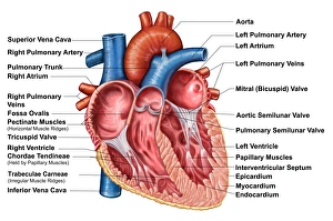
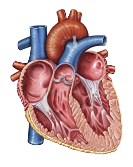
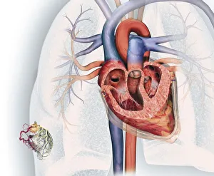
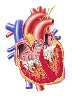
The left ventricle is a vital component of the heart's interior anatomy


All Professionally Made to Order for Quick Shipping
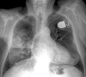
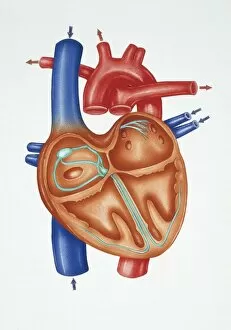
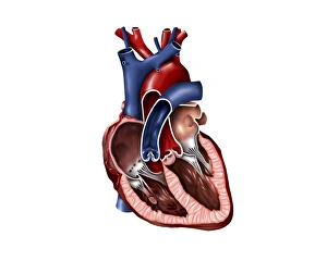
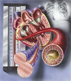
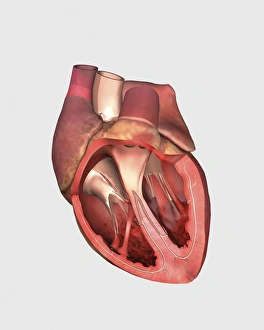
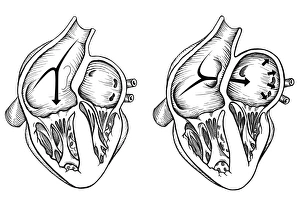
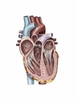
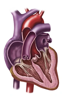
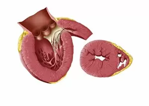
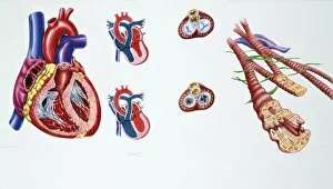
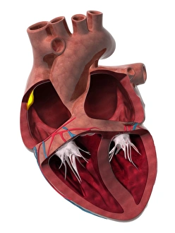
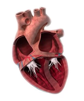
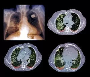
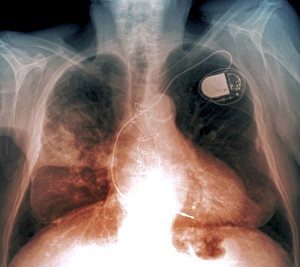
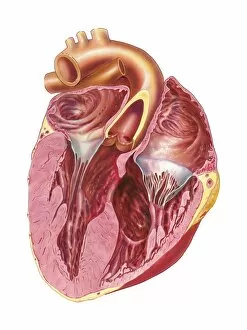
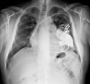
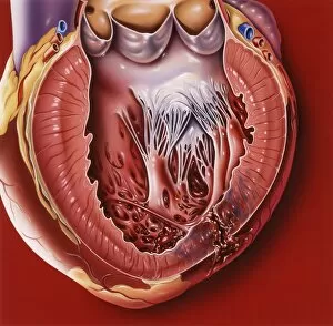
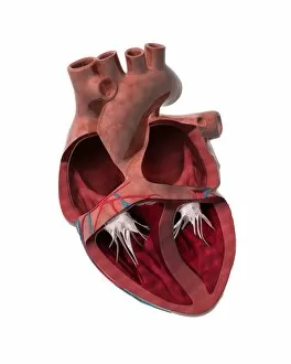
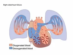
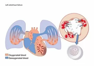
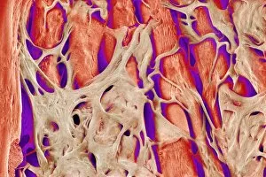
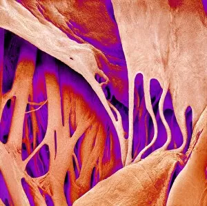
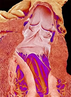
The left ventricle is a vital component of the heart's interior anatomy. In a frontal section, one can observe the intricate details of the human heart's interior, including the cross-section and the coronary system located at the bottom left. This system consists of essential elements such as the aorta, coronary arteries, coronary vein, and blood vessels. X-ray images depicting heart and lung diseases further emphasize the significance of understanding this crucial organ. By examining these X-rays (C018 / 0498), medical professionals gain valuable insights into diagnosing and treating various cardiac conditions. A closer look at a cross-section reveals more about the internal structure of our remarkable hearts. The muscle cells within are clearly visible along with an atherosclerotic artery - shedding light on potential cardiovascular issues caused by plaque buildup. Heart valves play an integral role in maintaining proper blood flow throughout our bodies. The pulmonary valve, mitral valve, and tricuspid valve are all showcased here to highlight their importance in ensuring efficient circulation. Comparing normal hearts to those with patent foramen ovale showcases how abnormalities can affect this complex organ's functionality. Understanding these differences aids in providing appropriate treatment options for patients. Finally, exploring an internal view allows us to appreciate both atria and ventricles within our hearts' interiors. These chambers work harmoniously to pump oxygenated blood throughout our bodies while removing waste products simultaneously. Delving into the intricacies of the left ventricle provides invaluable knowledge about human cardiac anatomy that is indispensable for healthcare professionals striving to diagnose and treat various cardiovascular conditions effectively.