Home > Arts > Street art graffiti > Digital art > Digital paintings
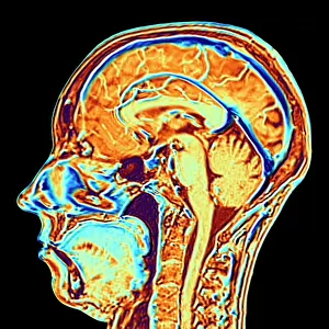
Digital illustration of parietal lobe (green), posterior superior temporal sulcas (blue), temperal pole (grey), dorsolateral prefrontal cortex, amygdala, and ventromedial prefrontal cortex (green) in human brain
, posterior superior temporal sulcas (blue), temperal pole (grey), dorsolateral prefrontal cortex, amygdala, and ventromedial prefrontal cortex (green) in human brain")
![]()
Wall Art and Photo Gifts from Fine Art Storehouse
Digital illustration of parietal lobe (green), posterior superior temporal sulcas (blue), temperal pole (grey), dorsolateral prefrontal cortex, amygdala, and ventromedial prefrontal cortex (green) in human brain
Unleash your creativity and transform your space into a visual masterpiece!
Dorling Kindersley
Media ID 13546359
© This content is subject to copyright
Amygdala Anatomy Biomedical Illustration Brain Brain Stem Cerebellum Cross Section Gray Green Grey Human Brain Parietal Lobe Physiology Square Square Image Digitally Generated Digitally Generated Image Green Color Human Body Part
FEATURES IN THESE COLLECTIONS
> Arts
> Street art graffiti
> Digital art
> Digital paintings
> Fine Art Storehouse
> Photo Libraries
> Dorling Kindersley Prints
> Fine Art Storehouse
> Science Inspired Art
> The Human Brain
> Fine Art Storehouse
> Science Inspired Art
EDITORS COMMENTS
This digital illustration by Dorling Kindersley showcases the intricate details of the human brain. The print features a cross-section view, revealing various regions and structures that make up this complex organ. In vibrant shades of green, we see the parietal lobe, dorsolateral prefrontal cortex, and ventromedial prefrontal cortex, highlighting their importance in cognitive functions and decision-making processes. The blue posterior superior temporal sulcas adds a touch of contrast to the composition while drawing attention to its role in auditory processing. The grey temporal pole stands out as a distinct feature, contributing to memory formation and emotional regulation within the brain. With no people present in this digitally generated image against a clean white background, our focus is solely on understanding the anatomy and physiology of these brain regions. This square format allows for an intimate exploration of each structure's unique characteristics. This biomedical illustration serves as both an educational tool for students studying neuroscience or medicine and an artistic representation of one of nature's most fascinating creations – the human brain. Its color palette brings life to what would otherwise be a clinical depiction, making it suitable for display in medical facilities or any setting where science meets artistry.
MADE IN THE USA
Safe Shipping with 30 Day Money Back Guarantee
FREE PERSONALISATION*
We are proud to offer a range of customisation features including Personalised Captions, Color Filters and Picture Zoom Tools
SECURE PAYMENTS
We happily accept a wide range of payment options so you can pay for the things you need in the way that is most convenient for you
* Options may vary by product and licensing agreement. Zoomed Pictures can be adjusted in the Cart.