Fraud Blocker Skip to main content

Parietal Lobe Collection

The parietal lobe, a vital region of the human brain, takes center stage in this captivating image

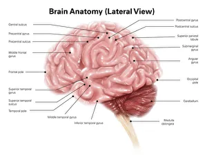
Background imageParietal Lobe Collection: Human brain anatomy, lateral view

Human brain anatomy, lateral view

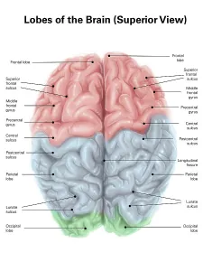
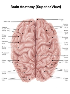
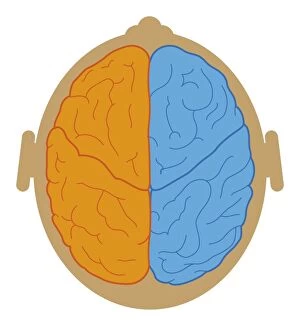
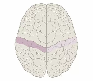
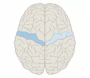
Background imageParietal Lobe Collection: Superior view of human brain with colored lobes and labels

Superior view of human brain with colored lobes and labels

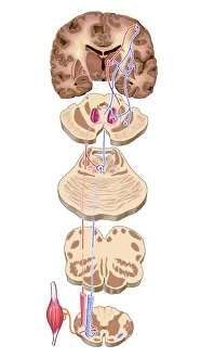
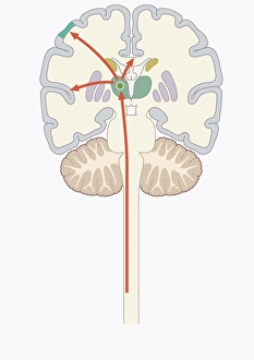
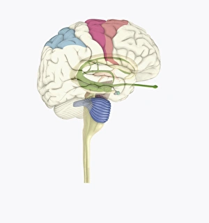
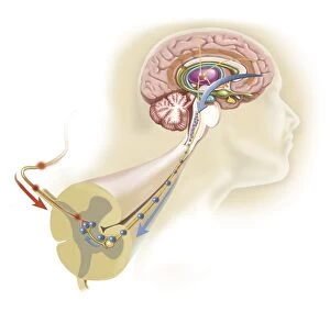
Background imageParietal Lobe Collection: Brain motor cortex pathways, artwork C016 / 6532

Brain motor cortex pathways, artwork C016 / 6532
Brain motor cortex pathways. Artwork of a sectioned human brain, brainstem and spinal cord, showing neural pathways (red and blue) from the motor cortex of the brain

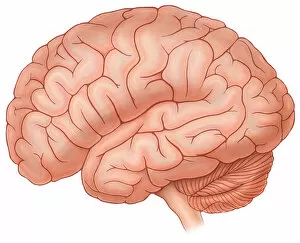
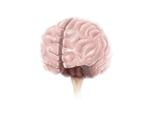
Background imageParietal Lobe Collection: Lateral view of a normal brain

Lateral view of a normal brain
Horizontal, Brain, Detail, Anatomy, Diagram, Illustration, Pink, Medical, Biology, Cut Out, White Background, Artwork, Color Image, Close Up, Healthcare And Medicine, The Human Body, No People

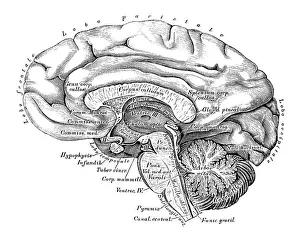
Background imageParietal Lobe Collection: Human anatomy scientific illustrations: Brain side view

Human anatomy scientific illustrations: Brain side view
Human anatomy scientific illustrations with latin/italian labels: Brain side view

Background imageParietal Lobe Collection: Human brain anatomy, superior view

Human brain anatomy, superior view

Background imageParietal Lobe Collection: Cross section biomedical illustration of brain map

Cross section biomedical illustration of brain map

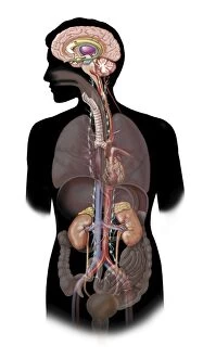
Background imageParietal Lobe Collection: Human body showing autonomic nervous system and limbic system

Human body showing autonomic nervous system and limbic system
Side view of human body showing autonomic nervous system and limbic system within the brain. Green are parasympathetic nerves. Blue are sympathetic nerves

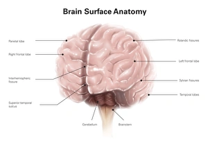
Background imageParietal Lobe Collection: Brain surface anatomy, with labels

Brain surface anatomy, with labels

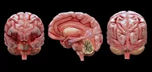
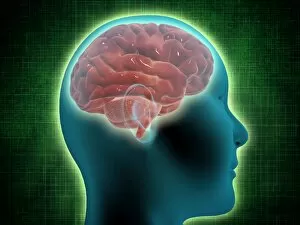
Background imageParietal Lobe Collection: 3D rendering of human brain

3D rendering of human brain

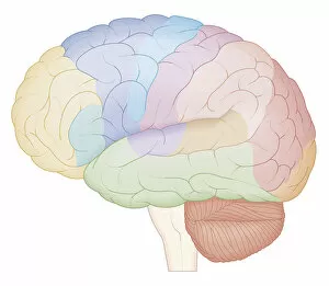
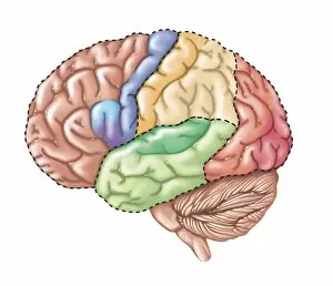
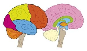
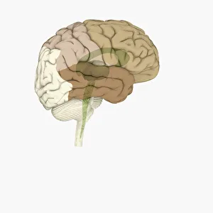
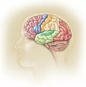
Background imageParietal Lobe Collection: Side view of the human brain showing the functional lobes

Side view of the human brain showing the functional lobes

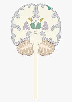
Background imageParietal Lobe Collection: Digital cross section illustration of human brain

Digital cross section illustration of human brain

Background imageParietal Lobe Collection: Digital illustration of pain signal to area of somatosensory cortex in human brain via spine

Digital illustration of pain signal to area of somatosensory cortex in human brain via spine

Background imageParietal Lobe Collection: Digital illustration of human brain showing lobes and cross section

Digital illustration of human brain showing lobes and cross section

Background imageParietal Lobe Collection: Brain anatomy

Brain anatomy
Illustration of a brain anatomy

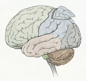
Background imageParietal Lobe Collection: Illustration of human brain showing frontal, temporal, occipital lobes

Illustration of human brain showing frontal, temporal, occipital lobes, section removed from parietal lobe, cerebellum and medulla oblongata

Background imageParietal Lobe Collection: Digital illustration of lobes and longitudinal fissure of human brain, close-up

Digital illustration of lobes and longitudinal fissure of human brain, close-up

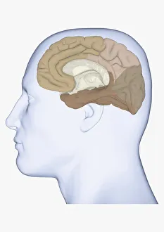
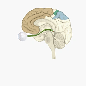
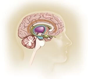
Background imageParietal Lobe Collection: Digital illustration of head in profile showing medial view of cortex in human brain

Digital illustration of head in profile showing medial view of cortex in human brain

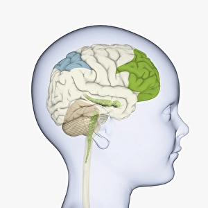
Background imageParietal Lobe Collection: Digital illustration of childs head in profile highlighting parts of brain

Digital illustration of childs head in profile highlighting parts of brain

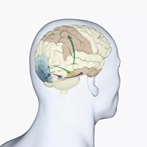
Background imageParietal Lobe Collection: Digital illustration of head in profile showing dorsal and ventral pathways of brain

Digital illustration of head in profile showing dorsal and ventral pathways of brain

Background imageParietal Lobe Collection: Digital illustration of highlighted areas in human brain affected by motor disorders

Digital illustration of highlighted areas in human brain affected by motor disorders

Background imageParietal Lobe Collection: Digital illustration of brain areas involved in altered states

Digital illustration of brain areas involved in altered states

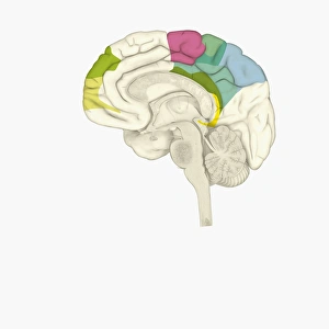
Background imageParietal Lobe Collection: Digital illustration of parts of human brain highlighted in shades of green, pink, blue, and yellow

Digital illustration of parts of human brain highlighted in shades of green, pink, blue, and yellow

Background imageParietal Lobe Collection: Digital illustration of head in profile showing lateral view of cortex in brain

Digital illustration of head in profile showing lateral view of cortex in brain

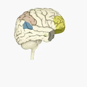
Background imageParietal Lobe Collection: Digital illustration of parietal lobe (green), posterior superior temporal sulcas (blue)

Digital illustration of parietal lobe (green), posterior superior temporal sulcas (blue), temperal pole (grey), dorsolateral prefrontal cortex, amygdala

Background imageParietal Lobe Collection: Digital illustration of various areas of cortex in human brain receiving input from sense organs

Digital illustration of various areas of cortex in human brain receiving input from sense organs

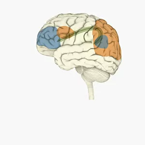
Background imageParietal Lobe Collection: Digital illustration of frontal lobe and parietal lobe areas (orange) in left hemispheres (blue)

Digital illustration of frontal lobe and parietal lobe areas (orange) in left hemispheres (blue), and pathway of data from parietal lobe to frontal lobe (green) in human brain

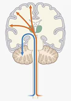
Background imageParietal Lobe Collection: Digital illustration of unconscious and conscious pathways in human brain passing through thalamus

Digital illustration of unconscious and conscious pathways in human brain passing through thalamus ending in parietal lobe of cortex

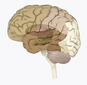
Background imageParietal Lobe Collection: Digital illustration of areas associated with memory in human brain

Digital illustration of areas associated with memory in human brain

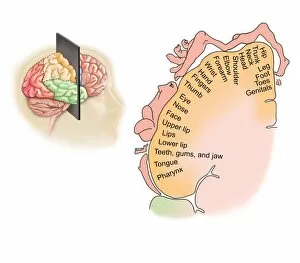
Background imageParietal Lobe Collection: Cross section biomedical illustration of touch map of human brain

Cross section biomedical illustration of touch map of human brain

Background imageParietal Lobe Collection: Cross section biomedical illustration of motor map of the brain

Cross section biomedical illustration of motor map of the brain

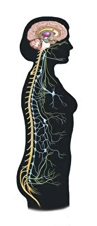
Background imageParietal Lobe Collection: Conceptual image of female body with brain, side view

Conceptual image of female body with brain, side view

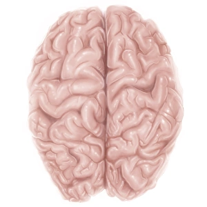
Background imageParietal Lobe Collection: Superior view of human brain

Superior view of human brain

Background imageParietal Lobe Collection: Brain surface anatomy

Brain surface anatomy

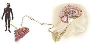
Background imageParietal Lobe Collection: Pathway of a pain message via sensory nerve in injured muscle

Pathway of a pain message via sensory nerve in injured muscle, to pain gate in spinal cord to limbic system, frontal cortex and sensory cortex in the human brain

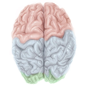
Background imageParietal Lobe Collection: Superior view of human brain with colored lobes

Superior view of human brain with colored lobes

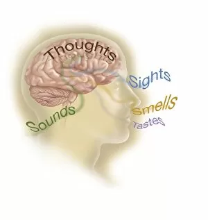
Background imageParietal Lobe Collection: Representation of how our senses affect our thoughts

Representation of how our senses affect our thoughts

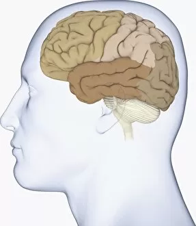
Background imageParietal Lobe Collection: Side view of the human head showing the functional lobes of the brain

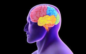
Side view of the human head showing the functional lobes of the brain
Side view of the human head showing the functional lobes of the human brain

Background imageParietal Lobe Collection: The sympathetic nervous system and the organs of fight-or-flight response

The sympathetic nervous system and the organs of fight-or-flight response

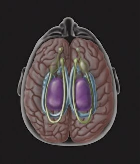
Background imageParietal Lobe Collection: View of limbic system as seen from directly above the head

View of limbic system as seen from directly above the head

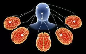
Background imageParietal Lobe Collection: Conceptual image of multi-brain processing

Conceptual image of multi-brain processing

Background imageParietal Lobe Collection: Human brain showing the layout of the sensory cortex

Human brain showing the layout of the sensory cortex
Coronal section through human brain showing the layout of the sensory cortex

Background imageParietal Lobe Collection: Anatomy of human brain, side view

Anatomy of human brain, side view

Background imageParietal Lobe Collection: Endorphins released in the hypothalamus travel down the spinal cord

Endorphins released in the hypothalamus travel down the spinal cord
Serotonin released in the brain travels down the spinal cord to close the pain gates and block pain messages

Background imageParietal Lobe Collection: Sagittal view of human brain showing the corpus callosum

Sagittal view of human brain showing the corpus callosum

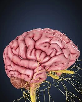
Background imageParietal Lobe Collection: Human brain with nerves

Human brain with nerves

Background imageParietal Lobe Collection: Side view of human brain showing internal carotid artery

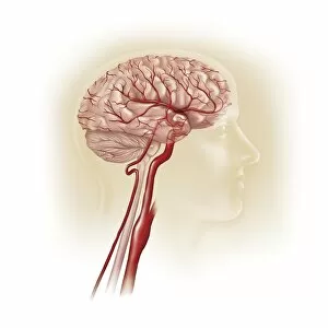
Side view of human brain showing internal carotid artery

Background imageParietal Lobe Collection: X-ray view of human face, profile view

X-ray view of human face, profile view


All Professionally Made to Order for Quick Shipping

The parietal lobe, a vital region of the human brain, takes center stage in this captivating image. With a superior view of the brain and its colored lobes clearly labeled, it offers an intriguing glimpse into our complex anatomy. This lateral view showcases the parietal lobe's position within the intricate network of neural pathways that make up our motor cortex. In another illustration, we are presented with a normal brain from a lateral perspective. Its parietal lobe stands out as one of the key players in our cognitive functions and sensory processing. The scientific precision of these illustrations allows us to appreciate the intricacies of human anatomy. A cross-section biomedical illustration further enhances our understanding by providing a detailed map of the brain's inner workings. It highlights how different regions interact, including those responsible for autonomic nervous system regulation and emotional responses found within the limbic system. As we shift to surface anatomy, labels guide us through each distinct feature on this side view of the human brain. Among them is none other than our star - the parietal lobe - which plays an essential role in spatial awareness, perception, attention, and language comprehension. Digital cross-section illustrations offer yet another perspective on this remarkable organ. They allow us to explore deeper layers and witness firsthand how various structures intertwine harmoniously to form such an intricate masterpiece. The parietal lobe truly shines amidst these captivating images showcasing its importance within our brains' functional lobes. As we delve into its complexities through scientific artistry and meticulous labeling, we gain profound insights into what makes us uniquely human –our ability to perceive ourselves and navigate through space with unparalleled cognition.