Fine Art Print : Skeletal muscle fibre


![]()
Fine Art Prints from Science Photo Library
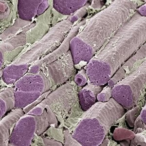
Skeletal muscle fibre
Skeletal muscle fibre. Coloured scanning electron micrograph (SEM) of skeletal muscle fibre. This type of muscle is striated. Muscle cells contain cylindrical organelles in the form of bundles of filaments (orange, myofibrils). Each filament can be thick (made up of myosin) or thin (made up of actin). In the case of skeletal (and cardiac) muscle, the filaments have a specific length, less than the length of the muscle cell. They are organised into repeating subunits (sarcomeres) along the length of the muscle cell. The resulting myofibrils run parallel to each other, causing the cell to appear striped (striated). The muscle is surrounded by connective tissue (loose strands)
Science Photo Library features Science and Medical images including photos and illustrations
Media ID 6448559
© SUSUMU NISHINAGA/SCIENCE PHOTO LIBRARY
Actin Connective Tissue Endomysium False Colour Fibre Fibres Filament Filaments Muscles Myofibril Myofibrils Myosin Orange Repeating Sarcomere Sarcomeres Skeletal Strand Strands Striated Striped Sub Unit Subunits Thick Thin Tissue False Coloured Repeat
20"x16" (+3" Border) Fine Art Print
Discover the intricacy of life with our Fine Art Prints from Media Storehouse. This captivating image showcases a coloured scanning electron micrograph (SEM) of a skeletal muscle fiber, revealing its intricate striated structure. A mesmerizing blend of science and art, this print brings the beauty of the human body to your home or office. Perfect for scientists, educators, or anyone with an appreciation for the wonders of biology. Experience the detail and vibrancy of this stunning print, a true conversation starter.
20x16 image printed on 26x22 Fine Art Rag Paper with 3" (76mm) white border. Our Fine Art Prints are printed on 300gsm 100% acid free, PH neutral paper with archival properties. This printing method is used by museums and art collections to exhibit photographs and art reproductions.
Our fine art prints are high-quality prints made using a paper called Photo Rag. This 100% cotton rag fibre paper is known for its exceptional image sharpness, rich colors, and high level of detail, making it a popular choice for professional photographers and artists. Photo rag paper is our clear recommendation for a fine art paper print. If you can afford to spend more on a higher quality paper, then Photo Rag is our clear recommendation for a fine art paper print.
Estimated Image Size (if not cropped) is 40.6cm x 50.8cm (16" x 20")
Estimated Product Size is 55.9cm x 66cm (22" x 26")
These are individually made so all sizes are approximate
Artwork printed orientated as per the preview above, with portrait (vertical) orientation to match the source image.
EDITORS COMMENTS
This print showcases the intricate details of a skeletal muscle fibre, captured using a scanning electron microscope (SEM). The image reveals the fascinating structure and composition of this type of muscle, which is known for its striated appearance. The muscle cells in the photo contain cylindrical organelles called myofibrils, represented by bundles of filaments in vibrant orange. These filaments can be thick or thin, composed respectively of myosin and actin proteins. Interestingly, these filaments have a specific length that is shorter than the overall length of the muscle cell. To create an organized pattern along the length of the muscle cell, these filaments are arranged into repeating subunits called sarcomeres. As a result, parallel myofibrils run alongside each other within the cell, giving it its characteristic striped or striated appearance. Surrounding this remarkable muscular structure is connective tissue depicted as loose strands in orange. This connective tissue provides support and protection to ensure proper functioning of the skeletal muscles. This stunning SEM image not only highlights the biological complexity but also serves as a testament to our ever-advancing understanding of anatomy and physiology. It offers viewers an opportunity to marvel at nature's design while gaining insight into how our bodies work on a microscopic level.
MADE IN THE USA
Safe Shipping with 30 Day Money Back Guarantee
FREE PERSONALISATION*
We are proud to offer a range of customisation features including Personalised Captions, Color Filters and Picture Zoom Tools
SECURE PAYMENTS
We happily accept a wide range of payment options so you can pay for the things you need in the way that is most convenient for you
* Options may vary by product and licensing agreement. Zoomed Pictures can be adjusted in the Cart.