
Actin Collection
"Actin: The Dynamic Force Behind Cell Structure and Muscle Contraction" From HeLa cells to muscle fibers, actin plays a crucial role in various biological processes


All Professionally Made to Order for Quick Shipping
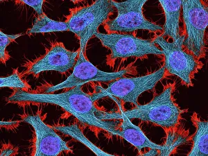
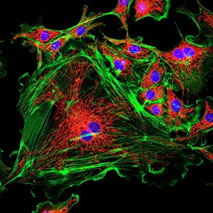
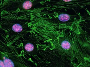
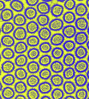
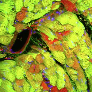
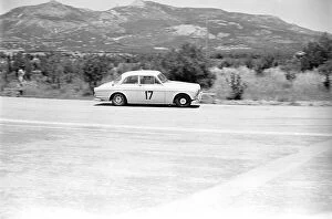
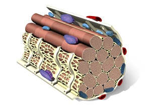
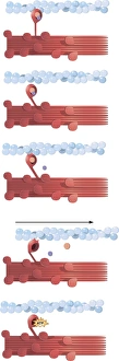
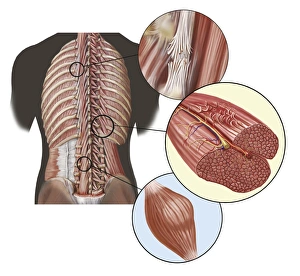
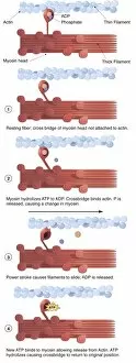
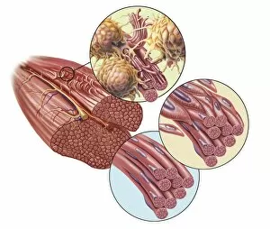
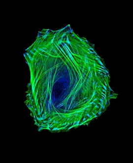
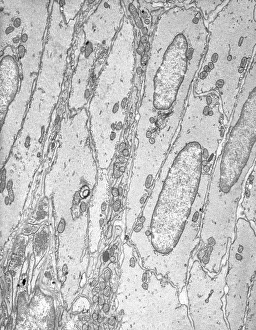
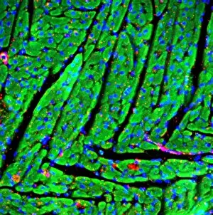
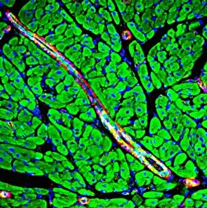
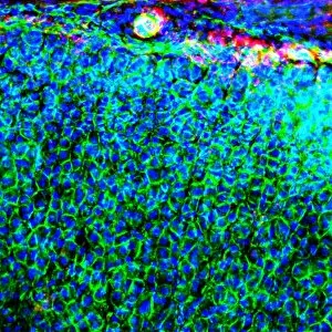
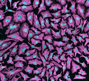
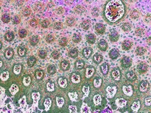
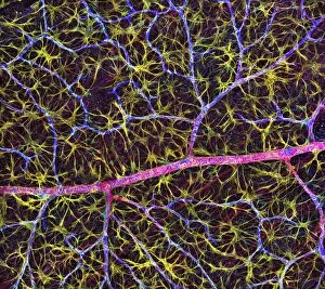
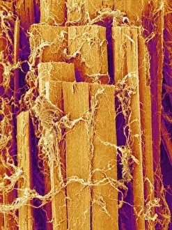
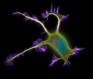
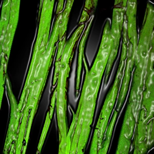
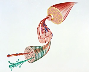
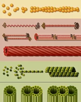
"Actin: The Dynamic Force Behind Cell Structure and Muscle Contraction" From HeLa cells to muscle fibers, actin plays a crucial role in various biological processes. This light micrograph C017 / 8299 showcases the intricate cell structure where actin filaments are prominently visible. In an immunofluorescent LM of fibroblast cell nuclei, actin's presence highlights its involvement in cellular movement and shape maintenance. Zooming into the intestinal microvilli through TEM reveals how actin filaments support their finger-like projections, aiding in nutrient absorption. Similarly, a confocal light micrograph of heart muscle unveils the organized arrangement and myosin filaments responsible for cardiac contractions. Stepping away from biology momentarily, we find ourselves at the 1973 24 Hours of Le Mans race. While seemingly unrelated, this event marked a breakthrough as researchers discovered similarities between car engines' pistons and muscle fiber structures - both relying on actin-myosin interactions for efficient performance. Returning to human anatomy, an artwork beautifully illustrates DTM (deep transverse massage) targeting deep back muscles affected by sprain, strain, or spasm. Actively engaging these muscles requires understanding their structure intimately. Delving deeper into muscle contraction mechanisms with labeled illustrations enlightens us about how actomyosin cross-bridges form during this process. Actins' ability to bind with myosins initiates muscular force generation necessary for any physical activity. Whether it's supporting cellular architecture or powering our movements during sports events like Le Mans or everyday activities like walking upstairs – "actin" remains an essential player behind the scenes.