Fine Art Print : Respiratory anatomy, 19th Century artwork

![]()
Fine Art Prints From Science Photo Library
Respiratory anatomy, 19th Century artwork
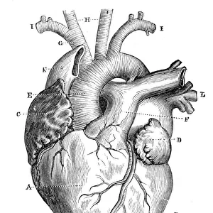
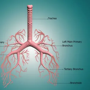
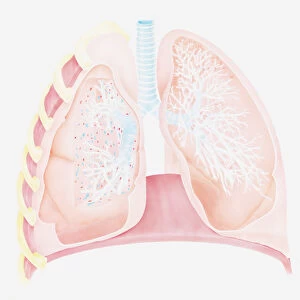
Respiratory anatomy, 19th Century artwork. Historical hand coloured lithographic print showing the trachea (wind pipe, vertical) running down from the larynx (voicebox, top centre) and branching (centre) into the two bronchi. These then branch further into numerous bronchioles inside each lung (left and right). Image from Traite complet de l anatomie de l homme, comprenant la medecine operatoire Vol. 4 (1836), by Jean-Baptiste Marc Bourgery and illustrated by Nicolas-Henri Jacob
Science Photo Library features Science and Medical images including photos and illustrations
Media ID 6327261
© SCIENCE PHOTO LIBRARY
1836 Airway Airways Bronchi Bronchiole Bronchioles Bronchus Chest Descriptive Anatomy Diagram French Frontal Interior Internal Larynx Lithograph Lithographic Print Lung Lungs Neck Organs Pulmonary Pulmonary System Respiratory System Thoracic Thorax Throat Trachea Vascular Vol 4 Volume Four Plate 6
20"x16" (+3" Border) Fine Art Print
Discover the captivating beauty of science's past with our Media Storehouse Fine Art Print of Respiratory Anatomy. This exquisite 19th-century artwork, sourced from Science Photo Library, offers a fascinating glimpse into the history of medical illustration. The intricately detailed hand-coloured lithographic print showcases the trachea, or windpipe, as it descends from the larynx, or voicebox. A stunning addition to any home or office, this fine art print not only serves as a beautiful decorative piece but also as a reminder of the rich history of scientific discovery. Bring a piece of history into your space and ignite your curiosity with this captivating work of art.
20x16 image printed on 26x22 Fine Art Rag Paper with 3" (76mm) white border. Our Fine Art Prints are printed on 300gsm 100% acid free, PH neutral paper with archival properties. This printing method is used by museums and art collections to exhibit photographs and art reproductions.
Our fine art prints are high-quality prints made using a paper called Photo Rag. This 100% cotton rag fibre paper is known for its exceptional image sharpness, rich colors, and high level of detail, making it a popular choice for professional photographers and artists. Photo rag paper is our clear recommendation for a fine art paper print. If you can afford to spend more on a higher quality paper, then Photo Rag is our clear recommendation for a fine art paper print.
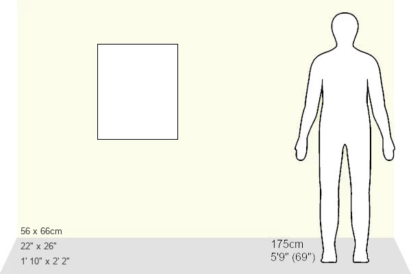
Estimated Image Size (if not cropped) is 40.6cm x 48.7cm (16" x 19.2")
Estimated Product Size is 55.9cm x 66cm (22" x 26")
These are individually made so all sizes are approximate
Artwork printed orientated as per the preview above, with portrait (vertical) orientation to match the source image.
EDITORS COMMENTS
This 19th-century artwork, a hand-coloured lithographic print titled "Respiratory Anatomy" offers a glimpse into the intricate structure of the human respiratory system. Created by Jean-Baptiste Marc Bourgery and illustrated by Nicolas-Henri Jacob, this historical piece showcases the internal workings of our lungs with remarkable detail. In this front view illustration set against a clean white background, we observe the trachea or windpipe descending vertically from the larynx at the top center. The trachea then branches out into two bronchi, which further divide into numerous bronchioles within each lung on either side. This depiction provides an insightful visual representation of how air travels through our respiratory system. The artist's skillful rendering highlights not only the anatomical accuracy but also captures the beauty inherent in biological structures. With its meticulous attention to detail and vibrant colors, this lithograph serves as both an artistic masterpiece and an educational tool for those interested in understanding human anatomy. Originally featured in "Traite complet de l'anatomie de l'homme" volume four published in 1836, this print holds significant historical value. It sheds light on early scientific advancements and contributes to our knowledge of pulmonary function. Science Photo Library proudly presents this extraordinary piece that combines artistry with scientific exploration—a testament to humanity's continuous quest for understanding our own bodies.
MADE IN THE USA
Safe Shipping with 30 Day Money Back Guarantee
FREE PERSONALISATION*
We are proud to offer a range of customisation features including Personalised Captions, Color Filters and Picture Zoom Tools
SECURE PAYMENTS
We happily accept a wide range of payment options so you can pay for the things you need in the way that is most convenient for you
* Options may vary by product and licensing agreement. Zoomed Pictures can be adjusted in the Basket.