
Larynx Collection
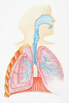
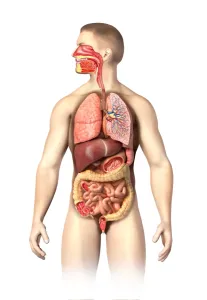
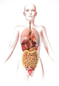
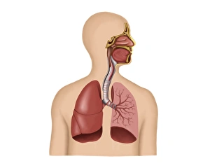
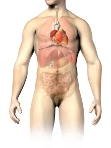
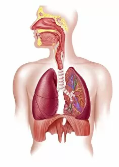
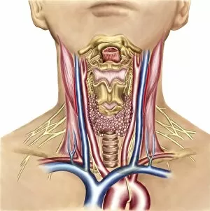
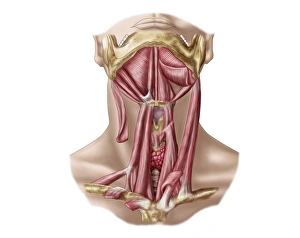
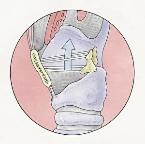
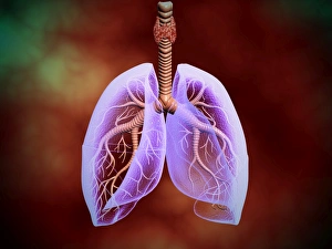
The larynx, also known as the voice box, is a vital part of the human respiratory system and can be seen in an illustration depicting the entire respiratory system


All Professionally Made to Order for Quick Shipping
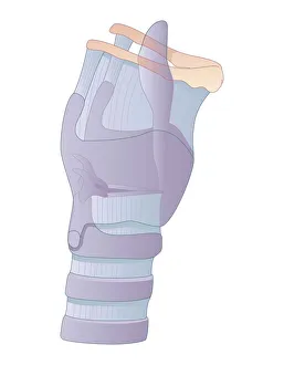
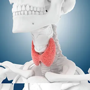
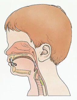
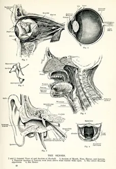
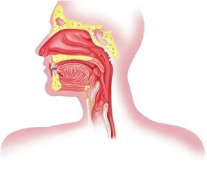
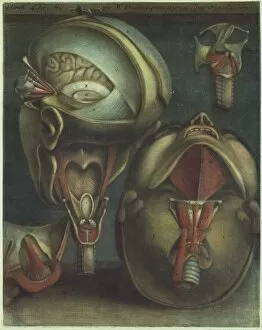
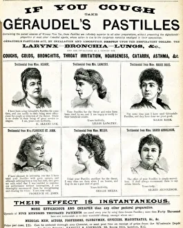
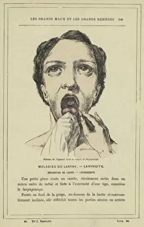
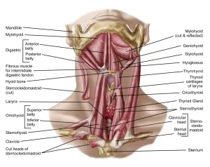
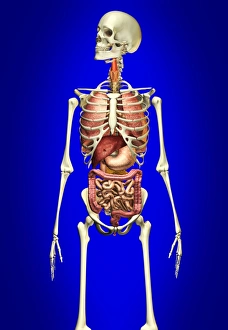
The larynx, also known as the voice box, is a vital part of the human respiratory system and can be seen in an illustration depicting the entire respiratory system, including the oral and nasal cavities, trachea, bronchus, and lungs. Another artwork showcases the intricate anatomy of the thyroid gland. Intriguingly, there is an engraving from 1866 that focuses on neck throat anatomy. This piece provides a detailed view of the larynx's position within our bodies. Furthermore, it highlights how this organ plays a crucial role in our ability to speak and produce sound. The importance of the larynx becomes even more apparent when considering its depiction alongside other senses in an artwork titled "Human Senses. " This emphasizes how our vocal abilities are intertwined with our perception of touch, taste, smell, sight, and hearing. Additionally, two artworks show different views of the head from 1746 by Jacques Fabien Gautier Dagoty. These pieces likely include representations of various internal organs such as the larynx. Interestingly enough, advertisements for Geraudels Pastilles may have utilized images or references to promote their product's benefits for soothing throat irritations related to conditions involving the larynx. Medical lithographs provide further insight into specific aspects related to this organ's examination and treatment. One lithograph demonstrates how a laryngoscope can be used to examine vocal apparatuses visually. Another vividly portrays invasive membranes affecting both throat and larynx during diphtheria or croup infections. Finally, a color lithograph shows a polyp being removed from someone's larynx using a similar instrument -the larnygoscope- highlighting medical interventions available for treating certain conditions affecting this delicate structure.