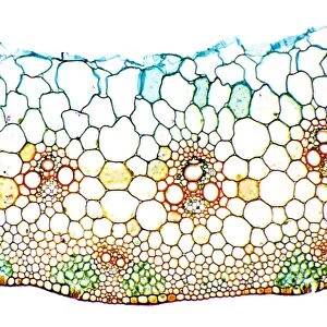
Wheat leaf, light micrograph

![]()
Wall Art and Photo Gifts from Science Photo Library
Wheat leaf, light micrograph
Wheat leaf. Light micrograph of a section through a leaf from a common wheat (Triticum aestivum) plant. The vascular bundle (centre to upper centre), or vein, of the leaf contains xylem vessels surrounded by xylem parenchyma (protoxylem). Underneath are larger vessels and smaller tracheids (red). beneath this is the phloem, which is composed of thin sieve tubes (red) and small companion cells. There are two outer circles around the vascular bundle, a pericycle (red) and bundle sheath (blue). The vein is joined to the lower epidermis by sclerenchyma supporting tissue. Magnification: x40 when printed 10 centimetres wide
Science Photo Library features Science and Medical images including photos and illustrations
Media ID 6354515
© DR KEITH WHEELER/SCIENCE PHOTO LIBRARY
Bundles Cell Biology Cytological Cytology Histological Histology Microscopy Monocot Monocots Monocotyledon Monocotyledons Parenchyma Pericycle Phloem Sieve Tube Sieve Tubes Stain Stained Structural Structures Tissue Tracheid Tracheids Vascular Bundle Vessels Xylem Vessel Cells Common Wheat Light Micrograph Light Microscope Section Sectioned Triticum Aestivum Vein
EDITORS COMMENTS
This print showcases the intricate structure of a wheat leaf, as seen through a light micrograph. The image reveals a section of a common wheat plant's leaf, providing an up-close look at its cellular composition. At the center and upper center of the leaf is the vascular bundle, or vein, which contains xylem vessels surrounded by xylem parenchyma known as protoxylem. Below this layer are larger vessels and smaller tracheids depicted in red. The phloem, responsible for transporting sugars throughout the plant, can be observed beneath these structures. It consists of thin sieve tubes also shown in red and small companion cells. Surrounding the vascular bundle are two outer circles: the pericycle portrayed in red and the bundle sheath represented in blue. To support its overall framework, sclerenchyma tissue connects the vein to the lower epidermis. This highly detailed image was captured using a light microscope with a magnification factor of 40 when printed at 10 centimeters wide. With its white background highlighting every cell and structure within it, this photograph provides valuable insights into botany and plant biology. Its stained appearance adds depth to our understanding of angiosperms' histology while emphasizing key components such as monocots' unique characteristics. Overall, this stunning visual representation serves as an invaluable resource for researchers studying plants' cellular makeup and their complex internal systems.
MADE IN THE USA
Safe Shipping with 30 Day Money Back Guarantee
FREE PERSONALISATION*
We are proud to offer a range of customisation features including Personalised Captions, Color Filters and Picture Zoom Tools
SECURE PAYMENTS
We happily accept a wide range of payment options so you can pay for the things you need in the way that is most convenient for you
* Options may vary by product and licensing agreement. Zoomed Pictures can be adjusted in the Cart.