Home > Popular Themes > Human Body
Osteoporosis

![]()
Wall Art and Photo Gifts from Science Photo Library
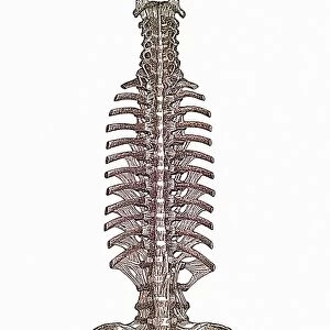
Osteoporosis
Osteoporosis. Computer artwork showing the anatomy of normal (left) and osteoporotic (right) bone. A bone shaft consists of an outer layer of compact bone made up of cylindrical units called osteons. This layer surrounds the honey-combed interior of spongy (cancellous) bone. In osteoporosis, it is the loss of the structural strength of spongy bone that causes brittle bones to snap. Across top, the sequence shows an osteoclast (green, a type of bone cell) destroying one of the linking supports of spongy bone tissue. Excess bone resorption can lead to osteoporosis. The osteoclast secretes acid granules (red, upper right) to dissolve the bone
Science Photo Library features Science and Medical images including photos and illustrations
Media ID 6415162
© HANS-ULRICH OSTERWALDER/SCIENCE PHOTO LIBRARY
Acid Bony Cancellous Compact Comparing Comparison Cut Away Destruction Diagram Diseased Dissolving Internal Nerve Nerves Osteoclast Osteocyte Osteological Osteology Osteon Osteons Osteoporosis Physiological Physiology Resorption Secreting Shaft Skeletal System Structural Tissue Trabecula Unit Vessels Computer Artwork Disorder Section
MADE IN THE USA
Safe Shipping with 30 Day Money Back Guarantee
FREE PERSONALISATION*
We are proud to offer a range of customisation features including Personalised Captions, Color Filters and Picture Zoom Tools
SECURE PAYMENTS
We happily accept a wide range of payment options so you can pay for the things you need in the way that is most convenient for you
* Options may vary by product and licensing agreement. Zoomed Pictures can be adjusted in the Cart.