Fraud Blocker Skip to main content

Home > Science > SEM

Embryonic stem cell and needle, SEM

 : Embryonic stem cell and needle, SEM
Sold 2
In Stock
We accept all major payment options so you can pay for the things you need in the way that is most convenient for you

Choose Your Product


Science Photo Library Photo Prints and Wall Art

Embryonic stem cell and needle, SEM

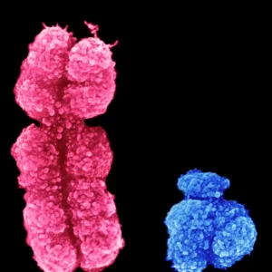
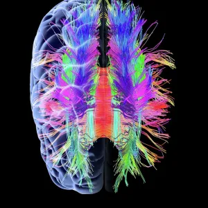
Embryonic stem cell and needle. Coloured scanning electron micrograph (SEM) of an embryonic stem cell (ESC) sitting in the eye of a needle. ESCs are pluripotent, that is they are able to differentiate into any cell type. The type of cell they mature into depends upon the biochemical signals received by the immature cells. This ability makes ESCs a potential source of cells to repair damaged tissue in diseases such as Parkinsons and insulin-dependent diabetes. However, research using ESCs is controversial as it requires the destruction of an embryo. Science Photo Library features Science and Medical images including photos and illustrations. © STEVE GSCHMEISSNER/SCIENCE PHOTO LIBRARY

Media ID 6399539

C Ulture Controversial Cultured Developmental Biology Embryonic Stem Cell Needle Pluripotent Precursor Cell Therapeutic Therapy Treatment Undifferentiated Cells False Coloured Genetics Micro Biology Scanning Electron Microscope Sewing Needle

FEATURES IN THESE COLLECTIONS

> Science > SEM

> Transportation > Cars > SIN

SECURE PAYMENTS
We accept all major payment options so you can pay for the things you need in the way that is most convenient for you

Made in USA
Safe Shipping, Easy Returns

FREE PERSONALISATION
We are proud to offer a range of customisation including Cropping, Captions, and Filters. Zoom Cropping can be adjusted in the basket


EDITORS COMMENTS

This print showcases the intricate world of embryonic stem cells. In this coloured scanning electron micrograph, we witness an embryonic stem cell delicately perched within the eye of a needle. These remarkable cells possess pluripotency, meaning they have the extraordinary ability to transform into any type of cell in our bodies. The potential held by these embryonic stem cells is immense. They offer hope for treating various diseases and conditions such as Parkinson's and insulin-dependent diabetes by replenishing damaged tissues. However, their use remains controversial due to the necessity of destroying embryos for research purposes. Through this image, we are reminded of the complex nature of biology and medicine. The vibrant colours highlight the beauty hidden within scientific exploration and discovery. It serves as a visual representation of cutting-edge concepts in developmental biology and microbiology. As we gaze upon this microscopic marvel, it becomes clear that these undifferentiated precursor cells hold tremendous promise for therapeutic applications in regenerative medicine. This photograph captures not only a single moment frozen in time but also represents a vast realm of possibilities waiting to be explored. Science Photo Library has once again provided us with an awe-inspiring glimpse into the wonders that lie beneath our very skin – reminding us that even at its smallest scale, life holds infinite potential for healing and growth.


Framed Prints of Embryonic stem cell and needle, SEM

Discover the wonders of scientific breakthroughs with our Media Storehouse Framed Prints. This captivating piece features an embryonic stem cell and needle in stunning detail, captured through the lens of Science Photo Library in a coloured scanning electron micrograph (SEM). Each print is meticulously framed to preserve the image's vibrant colours and intricate details, making it a mesmerizing addition to any laboratory, office, or home. Embrace the beauty of science and its endless discoveries with our Framed Prints.


Photo Prints of Embryonic stem cell and needle, SEM

Discover the wonders of scientific exploration with Media Storehouse's Photographic Prints. This captivating image, sourced from Science Photo Library, showcases an embryonic stem cell (ESC) in a coloured scanning electron micrograph (SEM). The ESC is delicately situated in the eye of a needle, a testament to the intricate and delicate nature of scientific research. Add this stunning print to your collection and bring the beauty of science into your home or office.


Poster Prints of Embryonic stem cell and needle, SEM

"Discover the wonders of scientific innovation with our Media Storehouse Poster Prints featuring the captivating image of an Embryonic Stem Cell and Needle, as captured in stunning detail through Scanning Electron Microscopy by Science Photo Library. This mesmerizing print showcases the intricate structure of an embryonic stem cell, poised at the tip of a needle, offering a glimpse into the groundbreaking realm of regenerative medicine. Elevate your workspace or learning environment with this thought-provoking and visually stimulating addition. Order your Media Storehouse Poster Print today and ignite curiosity and inspiration."


Jigsaw Puzzles of Embryonic stem cell and needle, SEM

Explore the wonders of science with Media Storehouse's Jigsaw Puzzles! This intriguing puzzle features an exquisite Colored Scanning Electron Micrograph (SEM) image from Science Photo Library, showcasing an Embryonic Stem Cell (ESC) delicately placed in the eye of a needle. This challenging puzzle is not just a fun activity, but an educational experience that brings the microscopic world to life. Engage your mind and sharpen your focus as you piece together the complex details of this remarkable image. Perfect for individuals of all ages and skill levels, our high-quality puzzles ensure hours of enjoyable and rewarding puzzling.

diamond
Made With High Grade Materials
thumb_up
Professional Quality Finish
package_2
Carefully Packed To Aid Safe Arrival

You May Also Like


Vascular bundle, SEMVascular bundle, SEM
Add to Basket
X and Y chromosomesX and Y chromosomes
Add to Basket
Northern lightsNorthern lights
Add to Basket
Mendeleyevs periodic table, 1869Mendeleyevs periodic table, 1869
Add to Basket
Self-portrait of Curiosity rover in Gale Crater on the surface of MarsSelf-portrait of Curiosity rover in Gale Crater on the surface of Mars
Add to Basket
Daltons table of Atomic symbols, 1835Daltons table of Atomic symbols, 1835
Add to Basket
Copper sulphate crystalsCopper sulphate crystals
Add to Basket
Rorschach Inkblot TestRorschach Inkblot Test
Add to Basket
Full body scan, MRI scanFull body scan, MRI scan
Add to Basket
Graphene sheet, artwork C016 / 8274Graphene sheet, artwork C016 / 8274
Add to Basket
White matter fibres and brain, artwork C015 / 1930White matter fibres and brain, artwork C015 / 1930
Add to Basket
US-APOLLO XI-RENDEZVOUSUS-APOLLO XI-RENDEZVOUS
Add to Basket

Gallery Wall Inspiration

Nerve cell, SEM
Red-barbed ant, SEM
Neutrophil engulfing MRSA, SEM C018 / 8596
Diatom alga, SEM
Diatom, SEM
X and Y chromosomes
Lavender pollen grain, SEM
Scanning Electron Micrograph (SEM): Tardigrade or ‘Water Bear Magnification x 1250 (A4 size: 29. 7 cm width)
Cat tongue surface, SEM
False-coloured SEM (Scanning Electron Micrograph) of a Tardigrade (Tardigrada) amongst moss, from Peak District National Park, Derbyshire, UK. This tardigrade is approximately 0. 1 millimetres long. Digital composite
Hazel pollen grains
Collagen, Scanning electron micrograph (SEM)
Santos-Dumont / Sem
T lymphocytes and cancer cell, SEM C001 / 1679
Tardigrade or Water Bear
Crysotile asbestos
Uterus lining during menstruation, SEM
Iris of the eye, SEM
Stylus in LP record
Salmonella bacteria, SEM
SEM of penicillin fungus
Vascular bundle, SEM
Geranium anther, SEM
Dahlia flower pollen, SEM
Scanning Electron Micrograph (SEM): Praying Mantis - Magnification x 30 (if print A4 size: 29. 7 cm wide)
Tardigrade, SEM
Scanning electron micrograph (SEM) of red blood cell


© Copyright Media Storehouse, All Rights Reserved 2003 - 2025
Facebook     Pinterest     100% Payment Secure     Reviews IO