Premium Framed Print : Human retina, 19th century artwork


![]()
Framed Photos From Science Photo Library
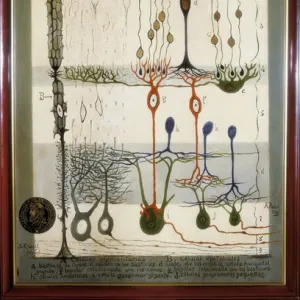
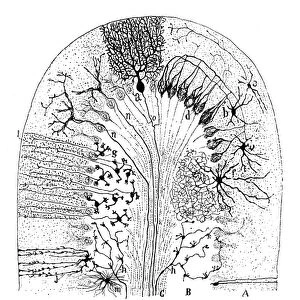
Human retina, 19th century artwork
Human retina, 19th century artwork. Artwork from the 1886 ninth edition of Moses and Geology (Samuel Kinns, London). This book was originally published in 1882
Science Photo Library features Science and Medical images including photos and illustrations
Media ID 6308781
© SCIENCE PHOTO LIBRARY
1882 1886 Diagram History Of Science Moses And Geology Ophthalmological Ophthalmology Retina Retinal Samuel Kinns Sense Sensory Sight Vision Cells Mono Chrome Neurological Neurology
14"x18" Premium Frame
Contemporary style Premium Wooden Frame with 8"x12" Print. Complete with 2" White Mat and 1.25" thick MDF frame. Printed on 260 gsm premium paper. Glazed with shatter proof UV coated acrylic glass. Backing is paper covered backing with rubber bumpers. Supplied ready to hang with a pre-installed sawtooth/wire hanger. Care Instructions: Spot clean with a damp cloth. Securely packaged in a clear plastic bag and envelope in a reinforced cardboard shipper
FSC Real Wood Frame and Double Mounted with White Conservation Mountboard - Professionally Made and Ready to Hang
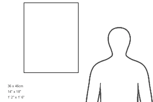
Estimated Image Size (if not cropped) is 20.3cm x 30.4cm (8" x 12")
Estimated Product Size is 35.6cm x 45.8cm (14" x 18")
These are individually made so all sizes are approximate
Artwork printed orientated as per the preview above, with portrait (vertical) orientation to match the source image.
EDITORS COMMENTS
This 19th-century artwork, titled "Human Retina" is a remarkable illustration from the ninth edition of Moses and Geology, published in 1886. Originally released in 1882 by Samuel Kinns in London, this historical masterpiece offers us a glimpse into the intricate world of human vision. Rendered with meticulous detail and presented in monochrome, this art piece showcases the delicate structure of our visual organ - the retina. The artist's skillful hand brings to life the complex network of cells that make up this vital component of our eyesight. As we delve into its anatomical intricacies, we are reminded of how far our understanding has come since these early scientific explorations. This illustration serves as a testament to the rich history of science and ophthalmology, shedding light on the foundations upon which modern neurology and biology have been built. Beyond its scientific significance, this artwork also possesses an undeniable aesthetic appeal. Its timeless beauty transcends time and invites us to appreciate both the artistic talent behind it and its historical context within medical illustration. Through this mesmerizing print from Science Photo Library, we are transported back to an era where knowledge was painstakingly documented through detailed illustrations like these – a true testament to humanity's unending quest for understanding ourselves and our world.
MADE IN THE USA
Safe Shipping with 30 Day Money Back Guarantee
FREE PERSONALISATION*
We are proud to offer a range of customisation features including Personalised Captions, Color Filters and Picture Zoom Tools
SECURE PAYMENTS
We happily accept a wide range of payment options so you can pay for the things you need in the way that is most convenient for you
* Options may vary by product and licensing agreement. Zoomed Pictures can be adjusted in the Basket.