Metal Print : Muscles of the foot

![]()
Metal Prints from Science Photo Library
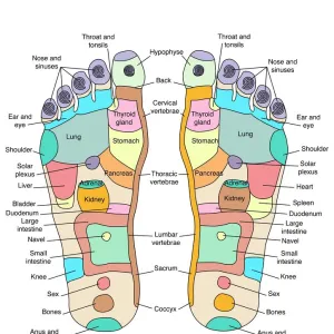
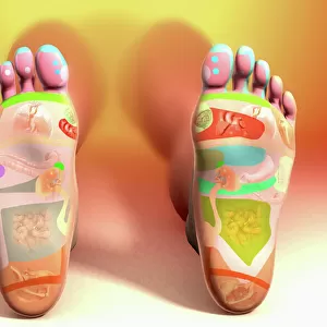
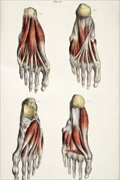
Muscles of the foot
Muscles of the foot, historical artwork. The figure at top left shows the first layer of muscles (red) in the sole of the foot. The skin and fascia (connective tissue) have been removed. At top right is the second layer, and at bottom right is the third and deepest layer of muscles in the sole of the foot. The underside of the heel bone (os calcis) is at top. Flexor and abductor muscles that move the toes attach to the heel bone. The figure at bottom left shows the muscles on the upper surface of the foot, which include the extensor muscles of the toes. Published in The Muscles of the Human Body... by Jones Quain in 1836
Science Photo Library features Science and Medical images including photos and illustrations
Media ID 6448613
© SHEILA TERRY/SCIENCE PHOTO LIBRARY
1836 19th Abductor Bones Carpal Tunnel Syndrome Dorsal Extensor Feet Flexor Foot Heel Historical Image Imagery Layer Layers Ligament Ligaments Muscle System Muscles Muscular Nineteenth Century Plane Planes Sole Tendon Tendons Toes Under Side Comments Jones Musculature Quain
16"x24" (61x41cm) Metal Print
Discover the intricacies of human anatomy with our stunning Metal Prints from Media Storehouse. This captivating artwork, originally published by AccuSoft Inc. in 1996-98 and sourced from Science Photo Library, showcases a detailed illustration of the muscles of the foot. With the top layer of muscles (in red) in the sole of the foot revealed, this historical piece offers a unique perspective into the complex structure of the foot. Bring this educational and aesthetically pleasing print into your home or office to inspire curiosity and ignite conversations. Order your Metal Print today and add a touch of science to your space.
Made with durable metal and luxurious printing techniques, our metal photo prints go beyond traditional canvases, adding a cool, modern touch to your space. Wall mount on back. Eco-friendly 100% post-consumer recycled ChromaLuxe aluminum surface. The thickness of the print is 0.045". Featuring a Scratch-resistant surface and Rounded corners. Backing hangers are attached to the back of the print and float the print 1/2-inch off the wall when hung, the choice of hanger may vary depending on size and International orders will come with Float Mount hangers only. Finished with a brilliant white high gloss surface for unsurpassed detail and vibrance. Printed using Dye-Sublimation and for best care we recommend a non-ammonia glass cleaner, water, or isopropyl (rubbing) alcohol to prevent harming the print surface. We recommend using a clean, lint-free cloth to wipe off the print. The ultra-hard surface is scratch-resistant, waterproof and weatherproof. Avoid direct sunlight exposure.
Made with durable metal and luxurious printing techniques, metal prints bring images to life and add a modern touch to any space
Estimated Product Size is 41.2cm x 61.5cm (16.2" x 24.2")
These are individually made so all sizes are approximate
Artwork printed orientated as per the preview above, with portrait (vertical) orientation to match the source image.
EDITORS COMMENTS
This historical artwork, titled "Muscles of the Foot" provides a detailed and intricate illustration of the muscular structure within our feet. Created by Jones Quain in 1836, this 19th-century print showcases three layers of muscles found in the sole of the foot. In the top left corner, we observe the first layer (depicted in red) after removing the skin and fascia. Moving to the top right, we encounter the second layer, followed by the third and deepest layer at bottom right. The underside of the heel bone (os calcis) is prominently displayed at the top, where flexor and abductor muscles responsible for toe movement attach. The figure at bottom left shifts our focus to explore muscles on the upper surface of our feet. This includes extensor muscles that play a crucial role in toe extension. With its meticulous attention to detail, this artwork offers valuable insights into foot anatomy from an earlier era. Published as part of "The Muscles of Human Body" series by Jones Quain, this image serves as a remarkable reference for anatomical study even today. It portrays various planes, tendons, ligaments while providing physiological references. With its rich imagery and comprehensive depiction of foot musculature dating back centuries ago, this historical image continues to be a valuable resource for those interested in understanding human anatomy or exploring medical history's artistic representations.
MADE IN THE USA
Safe Shipping with 30 Day Money Back Guarantee
FREE PERSONALISATION*
We are proud to offer a range of customisation features including Personalised Captions, Color Filters and Picture Zoom Tools
SECURE PAYMENTS
We happily accept a wide range of payment options so you can pay for the things you need in the way that is most convenient for you
* Options may vary by product and licensing agreement. Zoomed Pictures can be adjusted in the Cart.