Jigsaw Puzzle > Popular Themes > Human Body
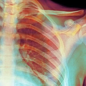
Jigsaw Puzzle : Tendinitis of the shoulder, X-ray


![]()
Jigsaw Puzzles From Science Photo Library
Tendinitis of the shoulder, X-ray
Tendinitis of the shoulder. Coloured X-ray of the shoulder of a patient showing calcification (grainy patches) around the rotator cuff tendon (upper left)
Science Photo Library features Science and Medical images including photos and illustrations
Media ID 6407007
© ZEPHYR/SCIENCE PHOTO LIBRARY
Calcification Human Body Part Injury Joint Medical Examination Orthopaedic Part Of The Body Radiography Rotator Cuff Shoulder Swelling Tendon Tendonitis X Ray X Ray Image Health Care Tendinitis
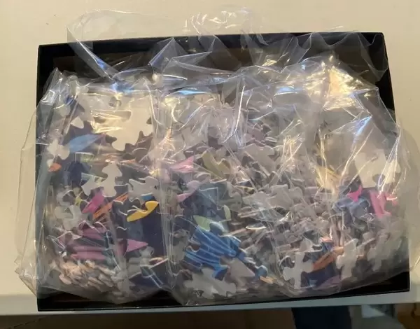
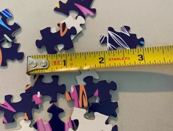
Jigsaw Puzzle (520 Pieces)
Discover the intricacies of the human body with our Media Storehouse Jigsaw Puzzles, featuring the captivating image "Tendinitis of the Shoulder, X-ray" from Science Photo Library. This puzzle offers an educational and engaging experience, allowing you to delve deep into the complexities of the shoulder joint. Explore the grainy calcifications around the rotator cuff tendon in vivid detail as you piece together this medical marvel. Challenge yourself and your family with this captivating and enlightening puzzle.
Made in the USA, 520-piece puzzles measure 16" x 20" (40.6 x 50.8 cm). Every puzzle is meticulously printed on glossy photo paper, which has a strong 1.33 mm thickness. Delivered in a black storage cardboard box, these puzzles are both stylish and practical. (Note: puzzles contain small parts and are not suitable for children under 3 years of age.)
Jigsaw Puzzles are an ideal gift for any occasion
Estimated Product Size is 40.5cm x 50.8cm (15.9" x 20")
These are individually made so all sizes are approximate
Artwork printed orientated as per the preview above, with landscape (horizontal) or portrait (vertical) orientation to match the source image.
EDITORS COMMENTS
This print from Science Photo Library showcases the intricate details of tendinitis in the shoulder. Against a striking black background, an X-ray image reveals the presence of calcification, depicted as grainy patches surrounding the rotator cuff tendon in the upper left region. The image provides valuable insights into this common injury that affects many individuals. Tendinitis occurs when tendons become inflamed due to repetitive motion or overuse, leading to pain and swelling. This visual representation allows us to observe how calcification can develop around the affected area, further complicating the condition. With its focus on biology and anatomy, this photograph serves as a powerful tool for medical professionals and researchers alike. By examining such detailed radiographic images like this one, orthopaedic specialists gain a deeper understanding of tendinitis and its impact on patients' health. As we explore this remarkable x-ray image, it is evident that our bodies are complex structures requiring proper care and attention. The rotator cuff plays a crucial role in maintaining shoulder joint stability and mobility; thus, any disruption caused by conditions like tendinitis can significantly affect daily activities. Science Photo Library continues to provide invaluable resources for medical examination purposes through their vast collection of high-quality visuals capturing various aspects of human health care. This particular print offers an enlightening glimpse into tendinitis pathology while reminding us of the importance of taking proactive measures to maintain our musculoskeletal well-being.
MADE IN THE USA
Safe Shipping with 30 Day Money Back Guarantee
FREE PERSONALISATION*
We are proud to offer a range of customisation features including Personalised Captions, Color Filters and Picture Zoom Tools
SECURE PAYMENTS
We happily accept a wide range of payment options so you can pay for the things you need in the way that is most convenient for you
* Options may vary by product and licensing agreement. Zoomed Pictures can be adjusted in the Basket.