Jigsaw Puzzle : Schistosome fluke, SEM


![]()
Jigsaw Puzzles From Science Photo Library
Schistosome fluke, SEM
Schistosome fluke. Coloured scanning electron micrograph (SEM) of a schistosome (Schistosoma sp.) fluke worm, a cause of schistosomiasis in humans. This parasitic trematode (flatworm) lives in the veins around the large intestine, anchoring itself to the wall of the blood vessel with several suckers (one seen at centre), and feeding on its hosts blood. The worms cause fever and abdominal pain, and the eggs produced by the female worm can form obstructions of tissues known as granulomas
Science Photo Library features Science and Medical images including photos and illustrations
Media ID 10249903
© AMI images
Bilharzia Colored Disease Causing Flatworm Fluke Mouth Parasite Parasitic Parasitism Pathogenic Platyhelminthes Schistosoma Mansoni Schistosome Schistosomiasis Sucker Trematode Worm Genetics Pathogen
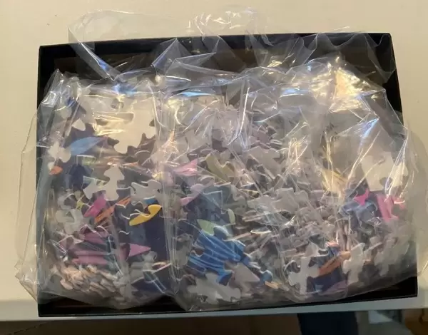
Jigsaw Puzzle (520 Pieces)
Discover the intricacies of microscopic life with Media Storehouse's Jigsaw Puzzles. Our latest addition to the collection showcases the mesmerizing Schistosome fluke in exquisite detail. This coloured Scanning Electron Micrograph (SEM) image by the National Cancer Institute from Science Photo Library brings the tiny yet complex world of Schistosoma sp. to your fingertips. Challenge yourself and your family with this captivating puzzle, perfect for both beginners and seasoned puzzle enthusiasts. Explore, learn, and connect the dots of science, one piece at a time.
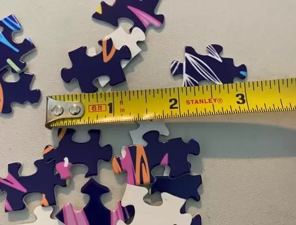
Made in the USA, 520-piece puzzles measure 16" x 20" (40.6 x 50.8 cm). Every puzzle is meticulously printed on glossy photo paper, which has a strong 1.33 mm thickness. Delivered in a black storage cardboard box, these puzzles are both stylish and practical. (Note: puzzles contain small parts and are not suitable for children under 3 years of age.)
Jigsaw Puzzles are an ideal gift for any occasion
Estimated Product Size is 40.5cm x 50.8cm (15.9" x 20")
These are individually made so all sizes are approximate
Artwork printed orientated as per the preview above, with landscape (horizontal) or portrait (vertical) orientation to match the source image.
EDITORS COMMENTS
This print showcases the intricate world of a Schistosome fluke, captured through a scanning electron microscope (SEM). These colored SEM images provide an astonishing glimpse into the life of this parasitic trematode, which is responsible for causing schistosomiasis in humans. The image reveals the adult worm's head and mouth, displaying its remarkable anatomy. The fluke worm resides within the veins surrounding the large intestine, firmly attaching itself to blood vessel walls using multiple suckers. It sustains its existence by feeding on its host's blood. Schistosomes inflict various symptoms upon their human hosts, including fever and abdominal pain. Additionally, eggs produced by female worms can lead to tissue obstructions called granulomas. This photograph serves as a powerful reminder of the devastating impact these creatures have on human health. With vibrant colors highlighting every detail, this SEM image not only provides valuable insights into biology and zoology but also serves as a diagnostic tool for identifying these disease-causing parasites. Its scientific significance lies in aiding researchers studying genetics and pathogenesis related to schistosomiasis. Through this visually stunning portrayal of nature's intricacies, we gain a deeper understanding of how such small organisms can have profound effects on our well-being.
MADE IN THE USA
Safe Shipping with 30 Day Money Back Guarantee
FREE PERSONALISATION*
We are proud to offer a range of customisation features including Personalised Captions, Color Filters and Picture Zoom Tools
SECURE PAYMENTS
We happily accept a wide range of payment options so you can pay for the things you need in the way that is most convenient for you
* Options may vary by product and licensing agreement. Zoomed Pictures can be adjusted in the Basket.