
Schistosomiasis Collection
Schistosomiasis, caused by Schistosoma spp. Blood flukes, is a parasitic disease that affects millions of people worldwide


All Professionally Made to Order for Quick Shipping
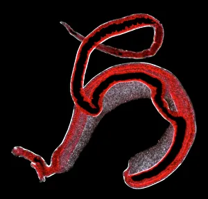
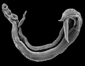
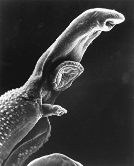
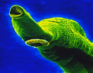
Schistosomiasis, caused by Schistosoma spp. Blood flukes, is a parasitic disease that affects millions of people worldwide. These microscopic parasites can penetrate the skin during contact with contaminated water, making it a significant public health concern in areas where sanitation and clean water access are limited. A scanning electron micrograph of a schistosome parasite reveals its intricate structure and highlights its ability to latch onto human hosts. The image showcases the complexity of these blood flukes and their adaptation for survival within the human body. Another micrograph captures the mating process of schistosome flukes, providing insight into their reproductive behavior. This fascinating glimpse into their life cycle sheds light on how they perpetuate their species within their host's bloodstream. Artwork depicting schistosome fluke worms further emphasizes the importance of understanding this disease. By visualizing these parasites, we gain a better understanding of their morphology and potential targets for intervention strategies. Colored SEM images showcase specific features of these blood flukes - from miracidium (the larval stage) to male head structures - enabling scientists to study them in greater detail. Such research aids in developing effective diagnostic tools and treatments against this debilitating disease. The SEM image revealing the mouth and sucker of the bilharzia fluke underscores its ability to attach itself firmly inside its host's body, causing damage to various organs over time if left untreated. This serves as a reminder that early detection is crucial for preventing severe complications associated with schistosomiasis. Finally, an LM image provides an overview of both male and female adult schistosome parasites within human blood vessels. Understanding their anatomy helps researchers identify potential vulnerabilities that could be targeted by drugs or vaccines aimed at eradicating this neglected tropical disease. Exploring different aspects through advanced imaging techniques offers valuable insights into this devastating illness caused by Schistosoma spp. Blood flukes.