Jigsaw Puzzle : Anatomy / Kidneys 18th C


![]()
Jigsaw Puzzles from Mary Evans Picture Library
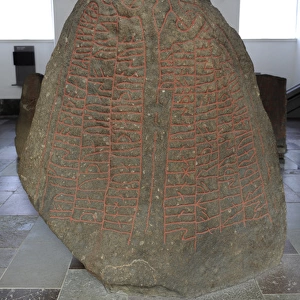
Anatomy / Kidneys 18th C
Anatomy of the kidneys and the intestines according to the anatomist Haller. Date: Circa 1760
Mary Evans Picture Library makes available wonderful images created for people to enjoy over the centuries
Media ID 7154893
© Mary Evans Picture Library 2015 - https://copyrighthub.org/s0/hub1/creation/maryevans/MaryEvansPictureID/10158228
1760 Anatomist Colon Haller Internal Intestine Intestines Kidney Kidneys Organ Organs
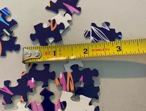
Jigsaw Puzzle (1014 Pieces)
Discover the fascinating world of human anatomy with our exquisite Jigsaw Puzzle from Media Storehouse, featuring the intricately detailed illustration "Anatomy / Kidneys 18th C" by Rights Managed from Mary Evans Prints Online. This captivating puzzle showcases an anatomical depiction of the kidneys and intestines as depicted by anatomist Haller around 1760. Immerse yourself in the history of medical discovery and challenge your mind with this intriguing and educational puzzle. Perfect for anatomists, history buffs, and puzzle enthusiasts alike.
Made in the USA, 1014-piece puzzles measure 20" x 30" (50.8 x 76.2 cm). Every puzzle is meticulously printed on glossy photo paper, which has a strong 1.33 mm thickness. Delivered in a black storage cardboard box, these puzzles are both stylish and practical. (Note: puzzles contain small parts and are not suitable for children under 3 years of age.)
Jigsaw Puzzles are an ideal gift for any occasion
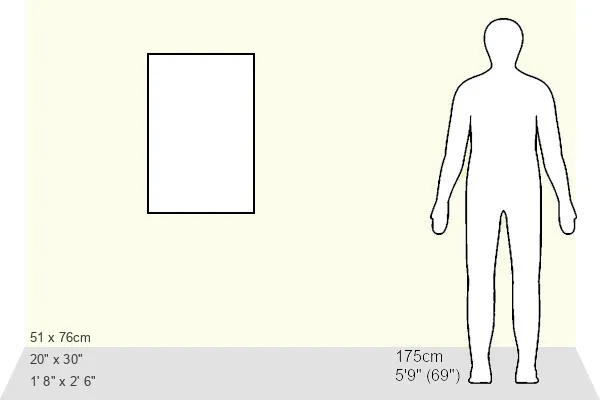
Estimated Product Size is 50.8cm x 76cm (20" x 29.9")
These are individually made so all sizes are approximate
Artwork printed orientated as per the preview above, with portrait (vertical) orientation to match the source image.
EDITORS COMMENTS
This stunning 18th century print depicts an intricate and detailed illustration of the anatomy of the kidneys and the intestines, as depicted by the renowned anatomist, Georg Heinrich von Haller. Dated circa 1760, this exquisite work of art provides a fascinating glimpse into the scientific understanding of the human body during this era. Haller, a Swiss physician and anatomist, was a leading figure in the field of anatomy during the Enlightenment period. His meticulous studies and illustrations contributed significantly to the advancement of medical knowledge, particularly in the areas of the urinary and digestive systems. The centerpiece of this print is the kidney, with its intricate structure of renal pyramids, calyces, and pelvis clearly visible. The ureter, which carries urine from the kidney to the bladder, is also depicted. Surrounding the kidney are the various structures of the urinary system, including the ureter, urinary bladder, and urethra. To the right of the kidney, the intestines and colon are illustrated in great detail. The large intestine, or colon, is shown with its various folds and structures, including the cecum, appendix, and ileocecal valve. The small intestine, with its villi and microvilli, is also depicted, highlighting the absorptive functions of this vital organ. This print offers a unique window into the scientific discoveries and advancements of the 18th century, providing a reminder of the rich history of medical illustration and the importance of anatomical studies in understanding the complex workings of the human body.
MADE IN THE USA
Safe Shipping with 30 Day Money Back Guarantee
FREE PERSONALISATION*
We are proud to offer a range of customisation features including Personalised Captions, Color Filters and Picture Zoom Tools
FREE COLORIZATION SERVICE
You can choose advanced AI Colorization for this picture at no extra charge!
SECURE PAYMENTS
We happily accept a wide range of payment options so you can pay for the things you need in the way that is most convenient for you
* Options may vary by product and licensing agreement. Zoomed Pictures can be adjusted in the Cart.