Fraud Blocker Skip to main content

Anatomist Collection

Anatomists have played a crucial role in unraveling the mysteries of the human body throughout history

Background imageAnatomist Collection: Shema histologique des nerfs de la colonne vertebrale. Dessin original en couleurs de Santiago

Shema histologique des nerfs de la colonne vertebrale. Dessin original en couleurs de Santiago Ramon y Cajal (1852-1934)
AIS5385609 Shema histologique des nerfs de la colonne vertebrale. Dessin original en couleurs de Santiago Ramon y Cajal (1852-1934), medecin histologiste espagnol, prix Nobel de medecine en 1906

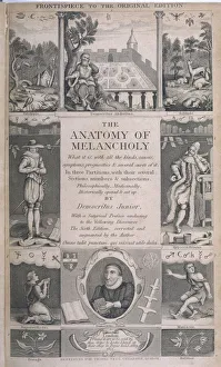
Background imageAnatomist Collection: Robert Burton

Robert Burton
ROBERT BURTON (1577-1640) English clergyman and author who wrote The Anatomy of Melancholy under the pseudonym of Democritus Junior in 1621

Background imageAnatomist Collection: Sir Charles Bell, 1824

Sir Charles Bell, 1824
1577003 Sir Charles Bell, 1824; (add.info.: Sir Charles Bell (12 November 1774 - 28 April 1842) was a Scottish surgeon and anatomist

Background imageAnatomist Collection: Portrait of Wilhelm Roux (1850-1924) German anatomist

Portrait of Wilhelm Roux (1850-1924) German anatomist
XEE5031760 Portrait of Wilhelm Roux (1850-1924) German anatomist; (add.info.: Portrait of Wilhelm Roux (1850-1924) German anatomist); Stefano Bianchetti

Background imageAnatomist Collection: The Sacred Heart of Jesus, 19th Century (colour litho)

The Sacred Heart of Jesus, 19th Century (colour litho)
942919 The Sacred Heart of Jesus, 19th Century (colour litho) by French School, (19th century); Private Collection; (add.info)

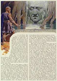
Background imageAnatomist Collection: Odin questioning Mimir (colour litho)

Odin questioning Mimir (colour litho)
7173279 Odin questioning Mimir (colour litho) by Doepler, Carl Emil (1824-1905); Private Collection; (add.info.: Odin questioning Mimir)

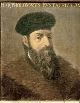
Background imageAnatomist Collection: EUSTACHIO, Bartolomeo (1500-1574). Italian anatomist

EUSTACHIO, Bartolomeo (1500-1574). Italian anatomist

Background imageAnatomist Collection: Portrait of a girl with a book, possibly Giulia di Alessandro de Medici, 1584-1550

Portrait of a girl with a book, possibly Giulia di Alessandro de Medici, 1584-1550, (oil on wood panel)
7146797 Portrait of a girl with a book, possibly Giulia di Alessandro de Medici, 1584-1550, (oil on wood panel) by Bronzino, Agnolo di Cosimo (1503-72); Galleria degli Uffizi, Florence, Tuscany

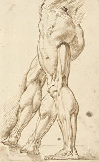
Background imageAnatomist Collection: An ecorche study of the legs of a male nude, with a subsidiary study of the right leg

An ecorche study of the legs of a male nude, with a subsidiary study of the right leg
3588687 An ecorchestudy of the legs of a male nude, with a subsidiary study of the right leg (pen & brown ink on paper) by Rubens

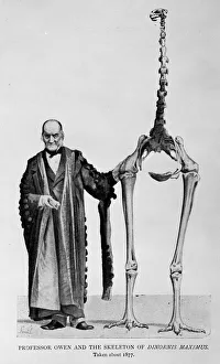
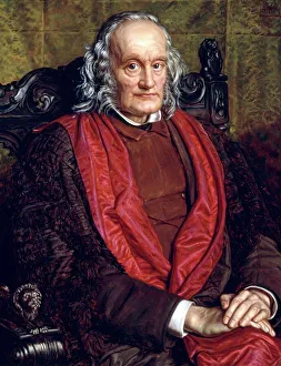
Background imageAnatomist Collection: Sir Richard Owen (1804-1892)

Sir Richard Owen (1804-1892)
Portrait of Sir Richard Owen, an English biologist, comparative anatomist and palaeontologist. Picture shows Owen and the skeleton of Dinornis maximus, c. 1877. From The Life of Owen (1894)

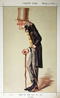
Background imageAnatomist Collection: 1873 Richard Owen Old bones Vanity Fair

1873 Richard Owen Old bones Vanity Fair
Richard Owen (20, July 1804- 18 December 1892). Caracature from Vanity Fairs Men of the Day series March 1st 1873. Owen was a comparative anatomist

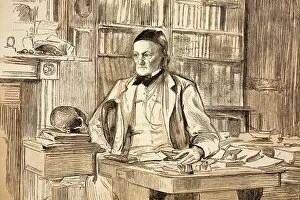
Background imageAnatomist Collection: 1883 Richard Owens study ex BMNH

1883 Richard Owens study ex BMNH
Richard Owen (20, July 1804- 18 December 1892). 1883 portrait in his home study for The Graphic magazine. The year he retired from the BMNH

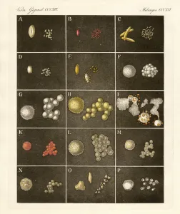
Background imageAnatomist Collection: The pollen or pollen of plants (coloured engraving)

The pollen or pollen of plants (coloured engraving)
998475 The pollen or pollen of plants (coloured engraving) by German School, (19th century); Private Collection; (add.info.: From A Bilderbuch fAor KinderA (Picture Book for Children) 1790-1830

Background imageAnatomist Collection: Richard Owen, British palaeontologist C016 / 5008

Richard Owen, British palaeontologist C016 / 5008
Richard Owen (1804-1892), British anatomist and palaeontologist. Owen was the founder and first director of the Natural History Museum, London, UK

Background imageAnatomist Collection: Guillaume Dupuytren

Guillaume Dupuytren
GUILLAUME DUPUYTREN French surgeon and anatomist

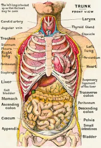
Background imageAnatomist Collection: Anatomical View of the Human Torso, 1914 (engraving)

Anatomical View of the Human Torso, 1914 (engraving)
2622096 Anatomical View of the Human Torso, 1914 (engraving) by German School, (20th century); Private Collection; (add.info.: Vintage anatomical study of the human torso, frontal view)

Background imageAnatomist Collection: Frontal View of Human Skull, 1917 (lithograph)

Frontal View of Human Skull, 1917 (lithograph)
2622467 Frontal View of Human Skull, 1917 (lithograph) by German School, (20th century); Private Collection; (add.info.: Vintage illustration of an anterior or frontal view of an anatomically correct)

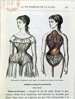
Background imageAnatomist Collection: The Harmful Effects of the Corset, illustration from La Vie Normale et la Sante

The Harmful Effects of the Corset, illustration from La Vie Normale et la Sante
CHT171318 The Harmful Effects of the Corset, illustration from La Vie Normale et la Sante by Dr Jules Rengade (b.1841) c.1880 (colour litho) by French School, (19th century); Bibliotheque Nationale

Background imageAnatomist Collection: Memorial to Francis Montagu Holl, English painter (photogravure)

Memorial to Francis Montagu Holl, English painter (photogravure)
7150716 Memorial to Francis Montagu Holl, English painter (photogravure) by Hollyer, Frederick (1837-1933) (after); Private Collection; (add.info.: Memorial to Francis Montagu Holl (1845-1888)

Background imageAnatomist Collection: Internal organs of the abdomen and thorax (colour litho)

Internal organs of the abdomen and thorax (colour litho)
3097100 Internal organs of the abdomen and thorax (colour litho) by German School, (19th century); Private Collection; (add.info.: Internal organs of the abdomen and thorax)

Background imageAnatomist Collection: Detail of a male anatomical ecorche model, displaying muscles, arteries and veins

Detail of a male anatomical ecorche model, displaying muscles, arteries and veins
CH469822 Detail of a male anatomical ecorche model, displaying muscles, arteries and veins, the circulation and nervous system (wax) (see also 469823 to 469827) by Zeiller

Background imageAnatomist Collection: Model of twins in the womb (wax)

Model of twins in the womb (wax)
BEN360190 Model of twins in the womb (wax) by Susini, Clemente (1754-1814); Museo della Specola, Florence, Italy; (add.info.: gemelli; scultura di cera; ); Photo eRaffaello Bencini; Italian

Background imageAnatomist Collection: Anatomical View of the Human Torso, 1914 (engraving)

Anatomical View of the Human Torso, 1914 (engraving)
2622098 Anatomical View of the Human Torso, 1914 (engraving) by German School, (20th century); Private Collection; (add.info.: Vintage anatomical study of the human torso, lateral view)

Background imageAnatomist Collection: Representation of an androgynous and a hermaphrodite. Plate taken from De natura Divinis

Representation of an androgynous and a hermaphrodite. Plate taken from De natura Divinis
GIA4791762 Representation of an androgynous and a hermaphrodite. Plate taken from De natura Divinis caracteris" by Cornelio Gemma 1575 by Unknown Artist

Background imageAnatomist Collection: Bones of the skeleton, from Tabulae Osteologicae

Bones of the skeleton, from Tabulae Osteologicae
CHT235091 Bones of the skeleton, from Tabulae Osteologicae by Christoph Jacob Trew (1695-1769) engraved by G. Lichtensteger, 1740 (coloured engraving) by Eisenberger

Background imageAnatomist Collection: Human anatomy. Ecorche by Jean Antoine Houdon (1741-1828), 18th century

Human anatomy. Ecorche by Jean Antoine Houdon (1741-1828), 18th century
JLJ4673665 Human anatomy. Ecorche by Jean Antoine Houdon (1741-1828), 18th century. Paris, School of Fine Arts by Houdon, Jean-Antoine (1741-1828); Bibliotheque de l Ecole des Beaux-Arts, Paris

Background imageAnatomist Collection: Surgical interventions on the female and male sexes. Engraving in '

Surgical interventions on the female and male sexes. Engraving in "
GIA4711054 Surgical interventions on the female and male sexes. Engraving in " Armamentarium Surgical" by J. Scultetus, Venice, 1665

Background imageAnatomist Collection: William Harvey explains the circulatory system to Charles I, using a live deer

William Harvey explains the circulatory system to Charles I, using a live deer, from El Museo Popular, 1889 (print)
693691 William Harvey explains the circulatory system to Charles I, using a live deer, from El Museo Popular, 1889 (print) by Spanish School, (19th century)

Background imageAnatomist Collection: Statue of John Hunter, by H. Weekes, R.A. in the Exhibition of the Royal Academy, 1864

Statue of John Hunter, by H. Weekes, R.A. in the Exhibition of the Royal Academy, 1864. Creator: Unknown
Statue of John Hunter, by H. Weekes, R.A. in the Exhibition of the Royal Academy, 1864. John Hunter...is, by common consent of his successors

Background imageAnatomist Collection: Karl Ernst Bock, 1809-1874 (engraving)

Karl Ernst Bock, 1809-1874 (engraving)
8632224 Karl Ernst Bock, 1809-1874 (engraving) by German School, (19th century) (after); Private Collection; (add.info.: Karl Ernst Bock, 1809-1874)

Background imageAnatomist Collection: Suspensory muscle of the duodenum.Small intestine, 1905 (engraving)

Suspensory muscle of the duodenum.Small intestine, 1905 (engraving)
8766911 Suspensory muscle of the duodenum.Small intestine, 1905 (engraving); Private Collection; (add.info.: Suspensory muscle of the duodenum.Small intestine. Human Anatomy by L)

Background imageAnatomist Collection: Cross section of a melanoid cancer, 1867 (engraving)

Cross section of a melanoid cancer, 1867 (engraving)
8766908 Cross section of a melanoid cancer, 1867 (engraving); Private Collection; (add.info.: Cross section of a melanoid cancer ( Dr. Jaccoud dictionary. Vol)

Background imageAnatomist Collection: Engraving of a pregnant woman's body, 1498, Johannes de Ketham

Engraving of a pregnant woman's body, 1498, Johannes de Ketham
AIS5390851 Engraving of a pregnant woman's body, 1498, Johannes de Ketham; Biblioteca Nacional, Madrid, Spain; (add.info.: Description of a pregnant woman's body)

Background imageAnatomist Collection: Breast, tongue, skin and finger sections. Lithograph of Corbie, based on a drawing by Leveille

Breast, tongue, skin and finger sections. Lithograph of Corbie, based on a drawing by Leveille
FLO4672795 Breast, tongue, skin and finger sections. Lithograph of Corbie, based on a drawing by Leveille, in Petity Atlas complet d'Anatomie descriptive du Corps Humain, by Dr

Background imageAnatomist Collection: Neck, tongue and throat muscles. Lithograph of Choubard, based on a drawing by Leveille

Neck, tongue and throat muscles. Lithograph of Choubard, based on a drawing by Leveille
FLO4672612 Neck, tongue and throat muscles. Lithograph of Choubard, based on a drawing by Leveille, in Petity Atlas complet d'Anatomie descriptive du Corps Humain, by Dr

Background imageAnatomist Collection: Vertical sectionof the mucous membrane, 1905 (engraving)

Vertical sectionof the mucous membrane, 1905 (engraving)
8766912 Vertical sectionof the mucous membrane, 1905 (engraving); Private Collection; (add.info.: Vertical sectionof the mucous membrane, end of stomach and beginning of duodenum)

Background imageAnatomist Collection: Fish-skin helmet, c.1800-1900 (fish skin)

Fish-skin helmet, c.1800-1900 (fish skin)
7264988 Fish-skin helmet, c.1800-1900 (fish skin); Manchester Museum, The University of Manchester, England; (add.info.: Helmet made from a large Porcupine fish)

Background imageAnatomist Collection: Monstrum acephalon : Woodcut, Monstrum acephalon, monster without a head

Monstrum acephalon : Woodcut, Monstrum acephalon, monster without a head. From the 1642 book Monstrorum Historia by
UIG5327113 Monstrum acephalon : Woodcut, Monstrum acephalon, monster without a head. From the 1642 book Monstrorum Historia by Ulisse Aldrovandi (Bologna, 1522-1605)

Background imageAnatomist Collection: Aries Biceps : Renaissance woodcut Ram. From the 1642 book Monstrorum Historia by Ulisse

Aries Biceps : Renaissance woodcut Ram. From the 1642 book Monstrorum Historia by Ulisse Aldrovandi (Bologna)
UIG5327069 Aries Biceps : Renaissance woodcut Ram. From the 1642 book Monstrorum Historia by Ulisse Aldrovandi (Bologna, 1522-1605). He is considered the founder of modern Natural History

Background imageAnatomist Collection: Lupus Marinus Lupus Scythius : This creature looks a bit like a lion with a baboon's nose

Lupus Marinus Lupus Scythius : This creature looks a bit like a lion with a baboon's nose
UIG5327065 Lupus Marinus Lupus Scythius : This creature looks a bit like a lion with a baboon's nose and mane well-tamed by conditioner

Background imageAnatomist Collection: Harpy Secunda : Harpyae secunda icon, Egyptian harpy. Renaissance Woodcut monsters

Harpy Secunda : Harpyae secunda icon, Egyptian harpy. Renaissance Woodcut monsters
UIG5327064 Harpy Secunda : Harpyae secunda icon, Egyptian harpy. Renaissance Woodcut monsters. From the 1642 book Monstrorum Historia by Ulisse Aldrovandi (Bologna, 1522-1605)

Background imageAnatomist Collection: Puer Cornutus : Puer cornutus: a child with horns. From the 1642 book Monstrorum Historia by

Puer Cornutus : Puer cornutus: a child with horns. From the 1642 book Monstrorum Historia by Ulisse Aldrovandi
UIG5327158 Puer Cornutus : Puer cornutus: a child with horns. From the 1642 book Monstrorum Historia by Ulisse Aldrovandi (Bologna, 1522-1605). He is considered the founder of modern Natural History

Background imageAnatomist Collection: Sus Capite & Pedibus Humanis : Sus capite & pedibus humanis, pig with human head and hands

Sus Capite & Pedibus Humanis : Sus capite & pedibus humanis, pig with human head and hands
UIG5327153 Sus Capite & Pedibus Humanis : Sus capite & pedibus humanis, pig with human head and hands. From the 1642 book Monstrorum Historia by Ulisse Aldrovandi (Bologna, 1522-1605)

Background imageAnatomist Collection: Pullus Equinus Humana Facie : Horse with human face. From the 1642 book Monstrorum Historia by

Pullus Equinus Humana Facie : Horse with human face. From the 1642 book Monstrorum Historia by Ulisse Aldrovandi
UIG5327149 Pullus Equinus Humana Facie : Horse with human face. From the 1642 book Monstrorum Historia by Ulisse Aldrovandi (Bologna, 1522-1605)

Background imageAnatomist Collection: Four Beasts : Hypertrichosis (also called Ambras syndrome) is an abnormal amount of hair growth

Four Beasts : Hypertrichosis (also called Ambras syndrome) is an abnormal amount of hair growth over the body;
UIG5327174 Four Beasts : Hypertrichosis (also called Ambras syndrome) is an abnormal amount of hair growth over the body; extensive cases of hypertrichosis have informally been called werewolf

Background imageAnatomist Collection: Agnus capite monstrifico : Agnus capite monstrifico, lamb with monstrous head

Agnus capite monstrifico : Agnus capite monstrifico, lamb with monstrous head
UIG5327169 Agnus capite monstrifico : Agnus capite monstrifico, lamb with monstrous head. From the 1642 book Monstrorum Historia by Ulisse Aldrovandi (Bologna, 1522-1605)

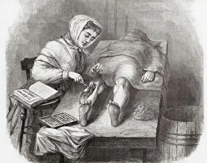
Background imageAnatomist Collection: Medical College for Women, New York

Medical College for Women, New York
7301631 Medical College for Women, New York.; Private Collection; (add.info.: A student dissecting a leg at the Medical College for Women on East Twelfth Street and Second Avenue, New York, USA)

Background imageAnatomist Collection: Anatomical section of the head

Anatomical section of the head
6266170 Anatomical section of the head; (add.info.: Anatomical section of the head. After a 19th century study by Robert Muys.); Photo © Ken Welsh. All rights reserved 2024

Background imageAnatomist Collection: Lo spellato (The bark) Bronze sculpture by Ludovico Cardi called Le Cigoli (1559-1613) Florence

Lo spellato (The bark) Bronze sculpture by Ludovico Cardi called Le Cigoli (1559-1613) Florence
BEN5479689 Lo spellato (The bark) Bronze sculpture by Ludovico Cardi called Le Cigoli (1559-1613) Florence. Museo Nazionale del Bargello by Cardi, Ludovico (1559-1613) ; Museo Nazionale del Bargello

Background imageAnatomist Collection: Lo spellato (The bark) Bronze sculpture by Ludovico Cardi called Le Cigoli (1559-1613) Florence

Lo spellato (The bark) Bronze sculpture by Ludovico Cardi called Le Cigoli (1559-1613) Florence
BEN5479860 Lo spellato (The bark) Bronze sculpture by Ludovico Cardi called Le Cigoli (1559-1613) Florence. Museo Nazionale del Bargello by Cardi, Ludovico (1559-1613) ; Museo Nazionale del Bargello

Background imageAnatomist Collection: Anatomy: knee joint Wax sculpture by Clemente Susini (1757-1814) Museo della Specola. florence

Anatomy: knee joint Wax sculpture by Clemente Susini (1757-1814) Museo della Specola. florence
BEN5478733 Anatomy: knee joint Wax sculpture by Clemente Susini (1757-1814) Museo della Specola. florence by Susini, Clemente (1754-1814); Photo © Raffaello Bencini

Background imageAnatomist Collection: Map of nerve bundles in the human brain by Paul Broca

Map of nerve bundles in the human brain by Paul Broca
5309460 Map of nerve bundles in the human brain by Paul Broca; (add.info.: Map of nerve bundles in the human brain by Paul Broca)

Background imageAnatomist Collection: Different views of the human heart

Different views of the human heart
5308008 Different views of the human heart.; (add.info.: Print depicting different views of the human heart by Bartolomeo Eustachi (d.1574) one of the founders of the science of human anatomy

Background imageAnatomist Collection: Octopus anatomy, ventral face, 1900 (engraving)

Octopus anatomy, ventral face, 1900 (engraving)
6947882 Octopus anatomy, ventral face, 1900 (engraving); Private Collection; (add.info.: Octopus anatomy, ventral face, 1900); Photo ©Jaime Abecasis

Background imageAnatomist Collection: Jaw of Lachesis muta (Grage Grand Carreau or master of the bush - venomous pit viper species jaw)

Jaw of Lachesis muta (Grage Grand Carreau or master of the bush - venomous pit viper species jaw)
CSO4996981 Jaw of Lachesis muta (Grage Grand Carreau or master of the bush - venomous pit viper species jaw), Museum d'histoire naturelle de Marseille; (add.info)

Background imageAnatomist Collection: Advertisement for Ashton Bennett Magnetic Corset Belts, 1923 (litho)

Advertisement for Ashton Bennett Magnetic Corset Belts, 1923 (litho)
8641318 Advertisement for Ashton Bennett Magnetic Corset Belts, 1923 (litho) by English School, (20th century); Private Collection; (add.info.: Advertisement for Ashton Bennett Magnetic Corset Belts)

Background imageAnatomist Collection: Human skeleton (coloured engraving)

Human skeleton (coloured engraving)
8655714 Human skeleton (coloured engraving) by French School, (19th century) (after); Private Collection; (add.info.: Human skeleton)

Background imageAnatomist Collection: Internal organs of a normal, healthy adult man (coloured engraving)

Internal organs of a normal, healthy adult man (coloured engraving)
8655763 Internal organs of a normal, healthy adult man (coloured engraving) by French School, (19th century) (after); Private Collection; (add.info.: Internal organs of a normal, healthy adult man)

Background imageAnatomist Collection: Intestinal absorption in the digestive system (coloured engraving)

Intestinal absorption in the digestive system (coloured engraving)
8655742 Intestinal absorption in the digestive system (coloured engraving) by French School, (19th century) (after); Private Collection; (add.info.: Intestinal absorption in the digestive system)

Background imageAnatomist Collection: Structure of the heart (coloured engraving)

Structure of the heart (coloured engraving)
8655735 Structure of the heart (coloured engraving) by French School, (19th century) (after); Private Collection; (add.info.: Structure of the heart)

Background imageAnatomist Collection: Nerves of the face (coloured engraving)

Nerves of the face (coloured engraving)
8655732 Nerves of the face (coloured engraving) by French School, (19th century) (after); Private Collection; (add.info.: Nerves of the face)

Background imageAnatomist Collection: Brain viewed in profile and cross-section (coloured engraving)

Brain viewed in profile and cross-section (coloured engraving)
8655728 Brain viewed in profile and cross-section (coloured engraving) by French School, (19th century) (after); Private Collection; (add.info.: Brain viewed in profile and cross-section)

Background imageAnatomist Collection: Connection of the sympathetic trunk with the vagus nerve and the spinal nerves (coloured engraving)

Connection of the sympathetic trunk with the vagus nerve and the spinal nerves (coloured engraving)
8655724 Connection of the sympathetic trunk with the vagus nerve and the spinal nerves (coloured engraving) by French School

Background imageAnatomist Collection: Evolution of the human skeleton (litho)

Evolution of the human skeleton (litho)
8672117 Evolution of the human skeleton (litho) by English School, (19th century); Private Collection; (add.info.: Evolution of the human skeleton, as described by Thomas Henry Huxley)

Background imageAnatomist Collection: Norma Lateralis (colour litho)

Norma Lateralis (colour litho)
8667626 Norma Lateralis (colour litho) by English School, (20th century); Private Collection; (add.info.: Norma Lateralis)

Background imageAnatomist Collection: Anterior View of Elbow Joint (colour litho)

Anterior View of Elbow Joint (colour litho)
8667622 Anterior View of Elbow Joint (colour litho) by English School, (20th century); Private Collection; (add.info.: Anterior View of Elbow Joint)

Background imageAnatomist Collection: Dissection of the Knee Joint from the Front, Patella thrown down (colour litho)

Dissection of the Knee Joint from the Front, Patella thrown down (colour litho)
8667620 Dissection of the Knee Joint from the Front, Patella thrown down (colour litho) by English School, (20th century); Private Collection; (add.info.: Dissection of the Knee Joint from the Front)

Background imageAnatomist Collection: The Muscles on the Back of the Arm, Forearm, and Hand (colour litho)

The Muscles on the Back of the Arm, Forearm, and Hand (colour litho)
8667619 The Muscles on the Back of the Arm, Forearm, and Hand (colour litho) by English School, (20th century); Private Collection; (add.info.: The Muscles on the Back of the Arm, Forearm, and Hand)

Background imageAnatomist Collection: Motor Points of the Upper Extremity, Anterior Aspect (litho)

Motor Points of the Upper Extremity, Anterior Aspect (litho)
8667614 Motor Points of the Upper Extremity, Anterior Aspect (litho) by English School, (20th century); Private Collection; (add.info.: Motor Points of the Upper Extremity, Anterior Aspect)

Background imageAnatomist Collection: Lynx lynx (Crane du lynx boreal ou lynx eurasian or wolf deer - Skull of an Eurasian lynx)

Lynx lynx (Crane du lynx boreal ou lynx eurasian or wolf deer - Skull of an Eurasian lynx)
CSO5006707 Lynx lynx (Crane du lynx boreal ou lynx eurasian or wolf deer - Skull of an Eurasian lynx), Museum d'histoire naturelle de Marseille; (add.info)

Background imageAnatomist Collection: engraving of the book of Ambroise Pare: Des Monsters et Prodiges (first edition 1573)

engraving of the book of Ambroise Pare: Des Monsters et Prodiges (first edition 1573)
PCT4260992 engraving of the book of Ambroise Pare: Des Monsters et Prodiges (first edition 1573, appendix: Monsters marine) - Triton et Sirene du Nile; (add.info)

Background imageAnatomist Collection: study of a body, hand, copperplate engraving, around 1700

study of a body, hand, copperplate engraving, around 1700
UTD4923684 study of a body, hand, copperplate engraving, around 1700; (add.info.: study of a body, hand, copperplate engraving)

Background imageAnatomist Collection: BARTHOLIN, Thomas (1616-1680). Danish physician, mathematician, and theologian

BARTHOLIN, Thomas (1616-1680). Danish physician, mathematician, and theologian
CUL4875538 BARTHOLIN, Thomas (1616-1680). Danish physician, mathematician, and theologian. He is best known for his work in the discovery of the lymphatic system in humans

Background imageAnatomist Collection: Human Anatomy book by MONTANA DE MONSERRATE, Bernardino, 1551 (engraving)

Human Anatomy book by MONTANA DE MONSERRATE, Bernardino, 1551 (engraving)
CUL4932051 Human Anatomy book by MONTANA DE MONSERRATE, Bernardino, 1551 (engraving) by Spanish School, (16th century); Biblioteca Nacional, Madrid, Spain; (add.info.: MONTANA DE MONSERRATE)

Background imageAnatomist Collection: Human Anatomy book by MONTANA DE MONSERRATE, Bernardino, 1551 (engraving)

Human Anatomy book by MONTANA DE MONSERRATE, Bernardino, 1551 (engraving)
CUL4932007 Human Anatomy book by MONTANA DE MONSERRATE, Bernardino, 1551 (engraving); Biblioteca Nacional, Madrid, Spain; (add.info.: MONTANA DE MONSERRATE, Bernardino (1480-1588). Spanish doctor

Background imageAnatomist Collection: Anatomical Plate - in ' De anatome corporis humani' by Guido Guidi (Vidus Vidius) (1509-1569)

Anatomical Plate - in " De anatome corporis humani" by Guido Guidi (Vidus Vidius) (1509-1569)
XEE5029671 Anatomical Plate - in " De anatome corporis humani" by Guido Guidi (Vidus Vidius) (1509-1569) Italian surgeon and anatomist

Background imageAnatomist Collection: Anatomical plate in ' De anatome corporis humani' by Guido Guidi (Vidus Vidius) (1509-1569)

Anatomical plate in " De anatome corporis humani" by Guido Guidi (Vidus Vidius) (1509-1569)
XEE5029670 Anatomical plate in " De anatome corporis humani" by Guido Guidi (Vidus Vidius) (1509-1569) Italian surgeon and anatomist

Background imageAnatomist Collection: Frontispice of ' De succo pancreatico', Leiden, 1671, by Regnier (Regnerus) de Graaf (1641 1673)

Frontispice of " De succo pancreatico", Leiden, 1671, by Regnier (Regnerus) de Graaf (1641 1673)
XEE5029587 Frontispice of " De succo pancreatico", Leiden, 1671, by Regnier (Regnerus) de Graaf (1641 1673) Dutch doctor - Engraving of 1672; (add.info)

Background imageAnatomist Collection: Monster creatures. Illustrated chart of a manuscript of Natural History by Ulisse Aldrovandi

Monster creatures. Illustrated chart of a manuscript of Natural History by Ulisse Aldrovandi (1522-1605)
VEN5029553 Monster creatures. Illustrated chart of a manuscript of Natural History by Ulisse Aldrovandi (1522-1605), Ulisse Aldrovandi fonds, University Library of Bologna, ed

Background imageAnatomist Collection: Monster creatures. Illustrated chart of a manuscript of Natural History by Ulisse Aldrovandi

Monster creatures. Illustrated chart of a manuscript of Natural History by Ulisse Aldrovandi (1522-1605)
VEN5029539 Monster creatures. Illustrated chart of a manuscript of Natural History by Ulisse Aldrovandi (1522-1605), Ulisse Aldrovandi fonds, University Library of Bologna, ed

Background imageAnatomist Collection: Monster creatures. Illustrated chart of a manuscript of Natural History by Ulisse Aldrovandi

Monster creatures. Illustrated chart of a manuscript of Natural History by Ulisse Aldrovandi (1522-1605)
VEN5029520 Monster creatures. Illustrated chart of a manuscript of Natural History by Ulisse Aldrovandi (1522-1605), Ulisse Aldrovandi fonds, University Library of Bologna, ed

Background imageAnatomist Collection: Cover page of ' Anatomia hepatis, cui praemittuntur quaedam ad rem anatomicam universe spectantia

Cover page of " Anatomia hepatis, cui praemittuntur quaedam ad rem anatomicam universe spectantia et ad calcem
XEE5029014 Cover page of " Anatomia hepatis, cui praemittuntur quaedam ad rem anatomicam universe spectantia et ad calcem operis subjiciuntur nonnulla de lymphae ductibus nuper repertis"

Background imageAnatomist Collection: Distribution of the hard portion of the auditory nerve - in ' Works Anatomiques'

Distribution of the hard portion of the auditory nerve - in " Works Anatomiques"
XEE5028966 Distribution of the hard portion of the auditory nerve - in " Works Anatomiques", Chez Charles-Antoine Jombert, 1761, Biblioteca nazionalez braidense


All Professionally Made to Order for Quick Shipping

Anatomists have played a crucial role in unraveling the mysteries of the human body throughout history. One such pioneer was Robert Burton, whose extensive research and writings on anatomy left an indelible mark on the field. Another notable figure was EUSTACHIO Bartolomeo, an Italian anatomist who made significant contributions during the 16th century. Intriguingly, a lithograph titled "Odin questioning Mimir" captures the essence of curiosity that drives anatomists to seek knowledge. This thirst for understanding is exemplified by Sir Richard Owen, a renowned British anatomist from the 19th century whose work continues to inspire modern scientists. Richard Owen's dedication to his craft is evident in various depictions of him throughout history. In one caricature from Vanity Fair in 1873, aptly titled "Old bones, " he is portrayed as a distinguished scholar with wisdom etched onto his face. An image from his study at BMNH in 1883 showcases his meticulous approach towards dissecting and studying specimens. The intricate details captured in engravings like "The Sacred Heart of Jesus" or "The pollen of plants" demonstrate how anatomists explore not only human anatomy but also delve into other aspects of nature's design. Richard Owen himself had a profound impact on paleontology, as depicted in C016 / 5008 - an image that pays homage to his groundbreaking discoveries. Throughout history, heart anatomists have held a special place within this scientific realm. Figures like Guillaume Dupuytren and William Hunter dedicated their lives to unraveling the complexities of this vital organ. Their contributions paved the way for advancements in cardiac medicine that continue to save lives today. From ancient times until now, these brilliant minds have shaped our understanding of ourselves and our world through their tireless pursuit of knowledge about our bodies' inner workings. The legacy they leave behind serves as an inspiration for future generations aspiring to unravel the secrets of human anatomy.

© Copyright Media Storehouse, All Rights Reserved 2003 - 2024
Facebook     Pinterest     Card Payments     Reviews IO