Fraud Blocker Skip to main content

Anatomist Collection (page 6)

Anatomists have played a crucial role in unraveling the mysteries of the human body throughout history

Background imageAnatomist Collection: Musculature in the male human body

Musculature in the male human body
6266188 Musculature in the male human body; (add.info.: Musculature in the male human body. After a 17th century work.); Photo © Ken Welsh. All rights reserved 2024.

Background imageAnatomist Collection: Octopus anatomy, ventral face, 1900 (engraving)

Octopus anatomy, ventral face, 1900 (engraving)
6947881 Octopus anatomy, ventral face, 1900 (engraving); Private Collection; (add.info.: Octopus anatomy , ventral face, 1900); Photo ©Jaime Abecasis.

Background imageAnatomist Collection: An engraving depicting a diagram showing the supposed functions of various areas of the brain

An engraving depicting a diagram showing the supposed functions of various areas of the brain, 19th century
5311364 An engraving depicting a diagram showing the supposed functions of various areas of the brain, 19th century; (add.info.: Date: 1886); Universal History Archive/UIG.

Background imageAnatomist Collection: A woodcut depicting twins in the uterus. Woodcut by Jost Amman (1539-1591) a Swiss-German artist

A woodcut depicting twins in the uterus. Woodcut by Jost Amman (1539-1591) a Swiss-German artist, 16th century
5311252 A woodcut depicting twins in the uterus. Woodcut by Jost Amman (1539-1591) a Swiss-German artist, 16th century; Universal History Archive/UIG.

Background imageAnatomist Collection: A human nervous system

A human nervous system
5308007 A human nervous system.; (add.info.: Print of a human nervous system by Bartolomeo Eustachi (d.1574) one of the founders of the science of human anatomy

Background imageAnatomist Collection: A human skeleton and nervous system

A human skeleton and nervous system
5308006 A human skeleton and nervous system.; (add.info.: Print of a human skeleton and nervous system by Bartolomeo Eustachi (d.1574) one of the founders of the science of human anatomy

Background imageAnatomist Collection: Merycoidodon culbertsoni (skull of an Oreodon (hillock teeth)

Merycoidodon culbertsoni (skull of an Oreodon (hillock teeth), Museum d'histoire naturelle de Marseille
CSO4987831 Merycoidodon culbertsoni (skull of an Oreodon (hillock teeth), Museum d'histoire naturelle de Marseille; (add.info.: Merycoidodon culbertsoni (skull of an Oreodon (hillock teeth)

Background imageAnatomist Collection: Crane de Palaeopropithecus sp (Madagascar lemurian, extirpated species - sloth lemur of)

Crane de Palaeopropithecus sp (Madagascar lemurian, extirpated species - sloth lemur of Madagascar, extinct)
CSO4987821 Crane de Palaeopropithecus sp (Madagascar lemurian, extirpated species - sloth lemur of Madagascar, extinct), Museum d'histoire naturelle de Marseille; (add.info)

Background imageAnatomist Collection: Megaladapis sp (Skullof an Extinct Mammalia from Madagascar) Museum d'histoire naturelle de

Megaladapis sp (Skullof an Extinct Mammalia from Madagascar) Museum d'histoire naturelle de Marseille
CSO4987812 Megaladapis sp (Skullof an Extinct Mammalia from Madagascar) Museum d'histoire naturelle de Marseille; (add.info)

Background imageAnatomist Collection: Medici Tigurini Historiae animalium

Medici Tigurini Historiae animalium
2626593 Medici Tigurini Historiae animalium; (add.info.: Illustration of a satyr or half man half beast monster, from 'medici Tigurini Historiae animalium' Historia animalium 1564; a)

Background imageAnatomist Collection: Medici Tigurini Historiae animalium

Medici Tigurini Historiae animalium
2626572 Medici Tigurini Historiae animalium; (add.info.: Illustration of a road runner bird, from 'medici Tigurini Historiae animalium' Historia animalium 1564; a veterinary anatomical book)

Background imageAnatomist Collection: Shi si jing fa hui

Shi si jing fa hui
2626557 Shi si jing fa hui; (add.info.: Illustration from the Japanese & Chinese edition (1716) of 'Shi si jing fa hui'

Background imageAnatomist Collection: Shi si jing fa hui

Shi si jing fa hui
2626545 Shi si jing fa hui; (add.info.: Illustration from the Japanese & Chinese edition (1716) of 'Shi si jing fa hui'

Background imageAnatomist Collection: Fasciculus medicinae

Fasciculus medicinae
2626479 Fasciculus medicinae; (add.info.: Illustration from 'Fasciculus medicinae' Venice; 1494 the first printed book to contain anatomical illustrations)

Background imageAnatomist Collection: A set of anatomical tables, with explanations, and an abridgment, of the practice of midwifery

A set of anatomical tables, with explanations, and an abridgment, of the practice of midwifery, 18th (engraving)
2626227 A set of anatomical tables, with explanations, and an abridgment, of the practice of midwifery, 18th (engraving); (add.info)

Background imageAnatomist Collection: A set of anatomical tables, with explanations, and an abridgment, of the practice of midwifery

A set of anatomical tables, with explanations, and an abridgment, of the practice of midwifery, 18th (engraving)
2626225 A set of anatomical tables, with explanations, and an abridgment, of the practice of midwifery, 18th (engraving); (add.info)

Background imageAnatomist Collection: Fasciculus medicinae

Fasciculus medicinae
2626473 Fasciculus medicinae; (add.info.: Illustration from 'Fasciculus medicinae' Venice; 1494 the first printed book to contain anatomical illustrations)

Background imageAnatomist Collection: Les oeuures d'Ambroise Pare?

Les oeuures d'Ambroise Pare?
2626428 Les oeuures d'Ambroise Pare?; (add.info.: Illustration from 'Les oeuures d'Ambroise Pare?' 1585 by Ambroise Pare)

Background imageAnatomist Collection: Equine anatomical Illustration

Equine anatomical Illustration
2626309 Equine anatomical Illustration; (add.info.: Equine anatomical Illustration from 'Anatomia del cavallo, infermita, et suoi rimedii')

Background imageAnatomist Collection: Fasciculus medicinae

Fasciculus medicinae
2626475 Fasciculus medicinae; (add.info.: Illustration from 'Fasciculus medicinae' Venice; 1494 the first printed book to contain anatomical illustrations)

Background imageAnatomist Collection: A set of anatomical tables, with explanations, and an abridgment, of the practice of midwifery

A set of anatomical tables, with explanations, and an abridgment, of the practice of midwifery, 18th (engraving)
2626221 A set of anatomical tables, with explanations, and an abridgment, of the practice of midwifery, 18th (engraving); (add.info)

Background imageAnatomist Collection: The anatomy textbook 'Ontleding des Menschelyken Lichaams', 1690 (engraving)

The anatomy textbook 'Ontleding des Menschelyken Lichaams', 1690 (engraving)
2625841 The anatomy textbook 'Ontleding des Menschelyken Lichaams', 1690 (engraving); (add.info.: Illustrations by Govard Bidloo)

Background imageAnatomist Collection: The Anatomy of Human Bodies, 1698 (engraving)

The Anatomy of Human Bodies, 1698 (engraving)
2625657 The Anatomy of Human Bodies, 1698 (engraving); (add.info.: Illustration from 'The anatomy of humane bodies' by William Cowper, (Oxford, 1698).); Universal History Archive/UIG.

Background imageAnatomist Collection: The Anatomy of Human Bodies, 1698 (engraving)

The Anatomy of Human Bodies, 1698 (engraving)
2625636 The Anatomy of Human Bodies, 1698 (engraving); (add.info.: Illustration from 'The anatomy of humane bodies' by William Cowper, (Oxford, 1698).); Universal History Archive/UIG.

Background imageAnatomist Collection: The Canon of Medicine

The Canon of Medicine
2625211 The Canon of Medicine; (add.info.: The central text consists of the anatomical sections of the Canon of Medicine by Ibn S?n? (Avicenna)

Background imageAnatomist Collection: Fetus head helped through the birth canal with forceps. Copperplate engraving by Andrew Bell after

Fetus head helped through the birth canal with forceps. Copperplate engraving by Andrew Bell after an illustration by
FLO4695943 Fetus head helped through the birth canal with forceps. Copperplate engraving by Andrew Bell after an illustration by Jan van Rymsdyk from William Smellie's A Set of Anatomical Tables

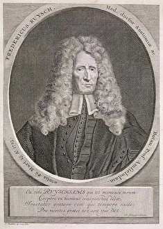
Background imageAnatomist Collection: Frederik Ruysch

Frederik Ruysch
2626864 Frederik Ruysch; (add.info.: Frederik Ruysch (1638-1731) anatomist. Dated 18th Century.); Universal History Archive/UIG.

Background imageAnatomist Collection: BARTHOLIN, Thomas (1616-1680). Danish physician, mathematician, and theologian

BARTHOLIN, Thomas (1616-1680). Danish physician, mathematician, and theologian
CUL4875561 BARTHOLIN, Thomas (1616-1680). Danish physician, mathematician, and theologian. He is best known for his work in the discovery of the lymphatic system in humans

Background imageAnatomist Collection: SUAREZ de RIBERA, Francisco (S.XVIII). Spanish doctor. Illustration of ' Theatro Chyrurgico

SUAREZ de RIBERA, Francisco (S.XVIII). Spanish doctor. Illustration of " Theatro Chyrurgico anatomico del cuerpo
CUL4871798 SUAREZ de RIBERA, Francisco (S.XVIII). Spanish doctor. Illustration of " Theatro Chyrurgico anatomico del cuerpo del hombre viviente

Background imageAnatomist Collection: SUAREZ de RIBERA, Francisco (S.XVIII). Spanish doctor. Illustration of ' Theatro Chyrurgico

SUAREZ de RIBERA, Francisco (S.XVIII). Spanish doctor. Illustration of " Theatro Chyrurgico anatomico del cuerpo
CUL4871759 SUAREZ de RIBERA, Francisco (S.XVIII). Spanish doctor. Illustration of " Theatro Chyrurgico anatomico del cuerpo del hombre viviente

Background imageAnatomist Collection: Le Coeur, extract from 'The anatomy of the human body' 1797, by Charles Bell (1774-1824)

Le Coeur, extract from "The anatomy of the human body" 1797, by Charles Bell (1774-1824)
XEE5028095 Le Coeur, extract from "The anatomy of the human body" 1797, by Charles Bell (1774-1824) - Biblioteca nazionale braidense, Milano; (add.info.: Le Coeur)

Background imageAnatomist Collection: Portrait of FELIX VICQ d'AZYR (1748-1794), French physician and anatomist

Portrait of FELIX VICQ d'AZYR (1748-1794), French physician and anatomist
XEE5032021 Portrait of FELIX VICQ d'AZYR (1748-1794), French physician and anatomist; (add.info.: Portrait of FELIX VICQ d'AZYR (1748-1794)

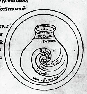
Background imageAnatomist Collection: Anatomy: representation of the uterus by Garioponto (970 - 1050), Italian doctor of the Middle Ages

Anatomy: representation of the uterus by Garioponto (970 - 1050), Italian doctor of the Middle Ages
XEE5029574 Anatomy: representation of the uterus by Garioponto (970 - 1050), Italian doctor of the Middle Ages, Biblioteca Civica Bertoliana

Background imageAnatomist Collection: Monster creatures. Illustrated chart of a manuscript of Natural History by Ulisse Aldrovandi

Monster creatures. Illustrated chart of a manuscript of Natural History by Ulisse Aldrovandi (1522-1605)
VEN5029555 Monster creatures. Illustrated chart of a manuscript of Natural History by Ulisse Aldrovandi (1522-1605), Ulisse Aldrovandi fonds, University Library of Bologna, ed

Background imageAnatomist Collection: Portrait of Antoine Ferrein (1693-1769) anatomist francais

Portrait of Antoine Ferrein (1693-1769) anatomist francais
XEE5029142 Portrait of Antoine Ferrein (1693-1769) anatomist francais; (add.info.: Portrait of Antoine Ferrein (1693-1769) anatomist francais); Stefano Bianchetti.

Background imageAnatomist Collection: Metoposcopiae liber, The metoposcopy of 1658, by (Cardanus Hieronymus) (Jerome Cardane)

Metoposcopiae liber, The metoposcopy of 1658, by (Cardanus Hieronymus) (Jerome Cardane) Gerolamo Cardano (1501-1576)
XEE5028461 Metoposcopiae liber, The metoposcopy of 1658, by (Cardanus Hieronymus) (Jerome Cardane) Gerolamo Cardano (1501-1576) Italian doctor, mathematician

Background imageAnatomist Collection: Bertillonage (Bertillon system or forensic anthropometry) a forensic technique developed by

Bertillonage (Bertillon system or forensic anthropometry) a forensic technique developed by the Frenchman Alphonse
ELD4961071 Bertillonage (Bertillon system or forensic anthropometry) a forensic technique developed by the Frenchman Alphonse Bertillon (1853-1914) in 1879

Background imageAnatomist Collection: The anatomist Niels Stensen (Stenon or Steensen or Steno or stenone) (1638-1688)

The anatomist Niels Stensen (Stenon or Steensen or Steno or stenone) (1638-1688)
ELD4905480 The anatomist Niels Stensen (Stenon or Steensen or Steno or stenone) (1638-1688) crowned of Titiopolis on 19/09/1677 by Pope Innocent XI and Cardinal Barbarigo

Background imageAnatomist Collection: France, Pays de la Loire, Maine-et-Loire (49), Angers: hospital of Angers, medical school

France, Pays de la Loire, Maine-et-Loire (49), Angers: hospital of Angers, medical school
LUX4771257 France, Pays de la Loire, Maine-et-Loire (49), Angers: hospital of Angers, medical school, medical students study anatomy with the help of a skeleton and the book

Background imageAnatomist Collection: External female parts of generation, labia, clitoris, perineum, and anus

External female parts of generation, labia, clitoris, perineum, and anus. Copperplate engraving by Andrew Bell after an
FLO4696243 External female parts of generation, labia, clitoris, perineum, and anus. Copperplate engraving by Andrew Bell after an illustration by Jan van Rymsdyk from William Smellie's A Set of

Background imageAnatomist Collection: Deep nervous system of the arm. Lithograph by Davesne, based on a drawing by Leveille

Deep nervous system of the arm. Lithograph by Davesne, based on a drawing by Leveille
FLO4673176 Deep nervous system of the arm. Lithograph by Davesne, based on a drawing by Leveille, in Petity Atlas complet d'Anatomie descriptive du Corps Humain, by Dr

Background imageAnatomist Collection: Brain, pons, spinal bulb, pineal gland etc... Lithograph by Davesne, based on a drawing by Leveille

Brain, pons, spinal bulb, pineal gland etc... Lithograph by Davesne, based on a drawing by Leveille
FLO4673157 Brain, pons, spinal bulb, pineal gland etc... Lithograph by Davesne, based on a drawing by Leveille, in Petity Atlas complet d'Anatomie descriptive du Corps Humain, by Dr

Background imageAnatomist Collection: Brachial plexus. Lithograph by Davesne, based on a drawing by Leveille

Brachial plexus. Lithograph by Davesne, based on a drawing by Leveille, in Petity Atlas complet d'Anatomie
FLO4673154 Brachial plexus. Lithograph by Davesne, based on a drawing by Leveille, in Petity Atlas complet d'Anatomie descriptive du Corps Humain, by Dr

Background imageAnatomist Collection: Section through the brain. Lithograph by Davesne, based on a drawing by Leveille

Section through the brain. Lithograph by Davesne, based on a drawing by Leveille
FLO4673112 Section through the brain. Lithograph by Davesne, based on a drawing by Leveille, in Petity Atlas complet d'Anatomie descriptive du Corps Humain, by Dr

Background imageAnatomist Collection: Different sections of the skull. Lithograph of Corbie, based on a drawing by Leveille

Different sections of the skull. Lithograph of Corbie, based on a drawing by Leveille
FLO4673087 Different sections of the skull. Lithograph of Corbie, based on a drawing by Leveille, in Petity Atlas complet d'Anatomie descriptive du Corps Humain, by Dr

Background imageAnatomist Collection: Lymph nodes and vessels of the head, neck and chest. Lithograph of Corbie

Lymph nodes and vessels of the head, neck and chest. Lithograph of Corbie, based on a drawing by Leveille
FLO4673058 Lymph nodes and vessels of the head, neck and chest. Lithograph of Corbie, based on a drawing by Leveille, in Petity Atlas complet d'Anatomie descriptive du Corps Humain, by Dr

Background imageAnatomist Collection: Lymph nodes and deep vessels of the leg. Lithograph of Corbie, based on a drawing by Leveille

Lymph nodes and deep vessels of the leg. Lithograph of Corbie, based on a drawing by Leveille
FLO4673028 Lymph nodes and deep vessels of the leg. Lithograph of Corbie, based on a drawing by Leveille, in Petity Atlas complet d'Anatomie descriptive du Corps Humain, by Dr

Background imageAnatomist Collection: Sinus or skull cavites. Humerus and vertebrae. Lithograph of Corbie, based on a drawing by Leveille

Sinus or skull cavites. Humerus and vertebrae. Lithograph of Corbie, based on a drawing by Leveille
FLO4673010 Sinus or skull cavites. Humerus and vertebrae. Lithograph of Corbie, based on a drawing by Leveille, in Petity Atlas complet d'Anatomie descriptive du Corps Humain, by Dr


All Professionally Made to Order for Quick Shipping

Anatomists have played a crucial role in unraveling the mysteries of the human body throughout history. One such pioneer was Robert Burton, whose extensive research and writings on anatomy left an indelible mark on the field. Another notable figure was EUSTACHIO Bartolomeo, an Italian anatomist who made significant contributions during the 16th century. Intriguingly, a lithograph titled "Odin questioning Mimir" captures the essence of curiosity that drives anatomists to seek knowledge. This thirst for understanding is exemplified by Sir Richard Owen, a renowned British anatomist from the 19th century whose work continues to inspire modern scientists. Richard Owen's dedication to his craft is evident in various depictions of him throughout history. In one caricature from Vanity Fair in 1873, aptly titled "Old bones, " he is portrayed as a distinguished scholar with wisdom etched onto his face. An image from his study at BMNH in 1883 showcases his meticulous approach towards dissecting and studying specimens. The intricate details captured in engravings like "The Sacred Heart of Jesus" or "The pollen of plants" demonstrate how anatomists explore not only human anatomy but also delve into other aspects of nature's design. Richard Owen himself had a profound impact on paleontology, as depicted in C016 / 5008 - an image that pays homage to his groundbreaking discoveries. Throughout history, heart anatomists have held a special place within this scientific realm. Figures like Guillaume Dupuytren and William Hunter dedicated their lives to unraveling the complexities of this vital organ. Their contributions paved the way for advancements in cardiac medicine that continue to save lives today. From ancient times until now, these brilliant minds have shaped our understanding of ourselves and our world through their tireless pursuit of knowledge about our bodies' inner workings. The legacy they leave behind serves as an inspiration for future generations aspiring to unravel the secrets of human anatomy.