
Spinal Collection (page 5)
"Unlocking the Secrets of Spinal Harmony: Exploring Chakras and the Nervous System" Delve into the intricate world of the spinal column


All Professionally Made to Order for Quick Shipping
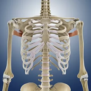
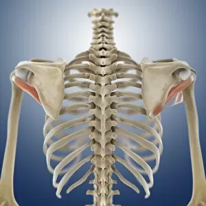
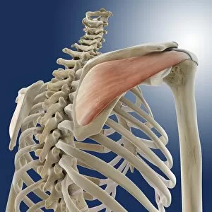
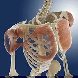
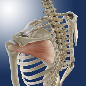
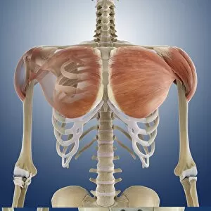
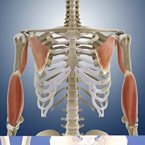
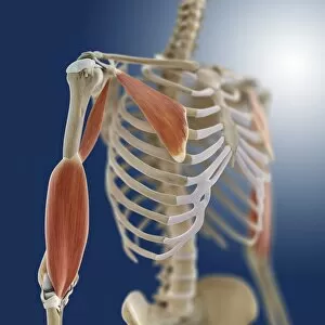
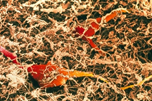
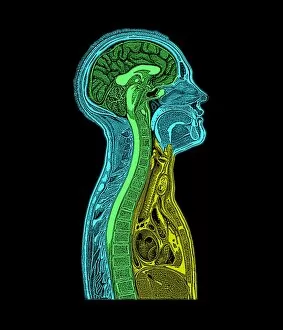
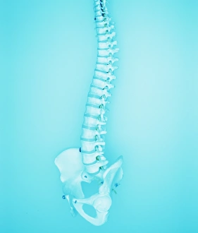
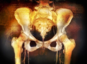
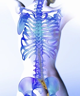
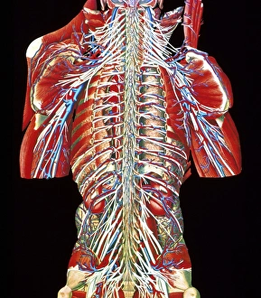
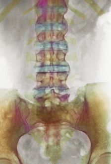
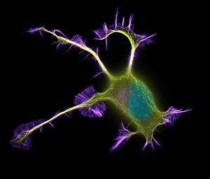
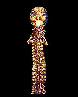
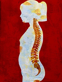
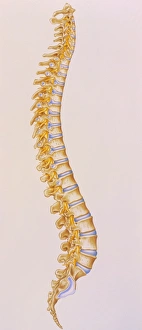
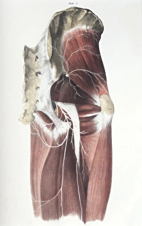
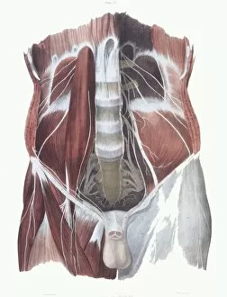
"Unlocking the Secrets of Spinal Harmony: Exploring Chakras and the Nervous System" Delve into the intricate world of the spinal column, where ancient wisdom meets modern science. Discover how this remarkable structure, revered by pioneers like Ramon y Cajal, holds the key to our physical and spiritual well-being. Embark on a visual journey through time as you explore a diagram showcasing the human brain and spinal cord. Witness how these interconnected powerhouses orchestrate every aspect of our existence, from bodily functions to emotional experiences. Step back in time with Calots spinal surgery, an innovation from the 19th century that revolutionized medical practices. Marvel at illustrations depicting this groundbreaking procedure that aimed to alleviate pain and restore mobility. Peer into X-ray images capturing both normal necks and spines, revealing their intricate beauty hidden beneath our skin. Observe an extended neck vertebrae X-ray, shedding light on potential issues caused by misalignment or injury. Art comes alive as body pain is transformed into captivating artwork – a poignant reminder of the challenges faced by those afflicted with discomfort along their spine. Let your imagination soar as upper body skeletons take form through computer-generated artistry. Witness how lower back pain manifests itself in conceptual artwork – a vivid representation of its impact on daily life. Explore similar expressions for upper back pain; each stroke conveying tales of struggle yet resilience against this common ailment. Intriguingly juxtaposing these artistic interpretations are X-rays showcasing normal necks and spines - silent testaments to health and balance within our bodies' core structures. Uncover new perspectives on spinal health as you immerse yourself in this captivating collection.