Fraud Blocker Skip to main content

Spinal Collection (page 4)

"Unlocking the Secrets of Spinal Harmony: Exploring Chakras and the Nervous System" Delve into the intricate world of the spinal column

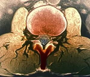
Background imageSpinal Collection: Healthy lumbar spine, MRI C018 / 0563

Healthy lumbar spine, MRI C018 / 0563
Healthy lumbar spine. Coloured magnetic resonance imaging (MRI) scan through the disc (red circle) between the second and third lumbar (lower back) vertebrae of a 30 year old patient

Background imageSpinal Collection: Healthy lumbar spine, MRI C018 / 0562

Healthy lumbar spine, MRI C018 / 0562
Healthy lumbar spine. Coloured magnetic resonance imaging (MRI) scan through the disc (purple) between the second and third lumbar (lower back) vertebrae of a 30 year old patient

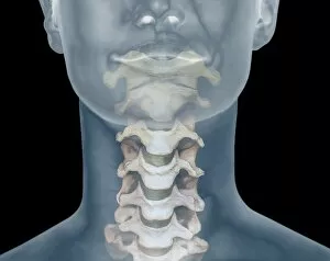
Background imageSpinal Collection: Healthy spine, CT scan C018 / 0444

Healthy spine, CT scan C018 / 0444
Healthy spine. Coloured 3D computed tomography (CT) scan of the cervical (neck) spine of a 28 year old. The spine is composed of blocks of bone called vertebrae

Background imageSpinal Collection: Healthy spine, CT scan C018 / 0441

Healthy spine, CT scan C018 / 0441
Healthy spine. Coloured 3D computed tomography (CT) scan of the cervical (neck) spine of a 28 year old. The spine is composed of blocks of bone called vertebrae

Background imageSpinal Collection: Spinal cord cyst, MRI scan C018 / 0561

Spinal cord cyst, MRI scan C018 / 0561
Spinal cord cyst. Coloured magnetic resonance imaging (MRI) scan of the cervical (neck) spinal cord of a 28 year old patient with syringomyelia, a cyst in the spinal cord

Background imageSpinal Collection: Healthy spine, CT scan C018 / 0442

Healthy spine, CT scan C018 / 0442
Healthy spine. Coloured 3D computed tomography (CT) scan of the cervical (neck) spine of a 28 year old. The spine is composed of blocks of bone called vertebrae

Background imageSpinal Collection: Osteoarthritis of the thoracic spine C017 / 0702

Osteoarthritis of the thoracic spine C017 / 0702
Osteoarthritis. False colour x-ray of the thorarcic spine of a 64 year old man with osteoarthritis. The arthritic vertebrae are closer together and have rough and ragged edges

Background imageSpinal Collection: Scoliosis treatment, X-ray C017 / 7156

Scoliosis treatment, X-ray C017 / 7156
Scoliosis treatment. X-ray of the spine of a 46 year old female patient that has been fixed to treat scoliosis (sideways curvature)

Background imageSpinal Collection: Hip joint bones and anatomy, artwork C014 / 2031

Hip joint bones and anatomy, artwork C014 / 2031
Hip joint bones and anatomy. Artwork of the bones that form the two hip joints (right and left). The hip is a ball-and-socket joint that provides the mobility needed for walking and running

Background imageSpinal Collection: Human spine, anatomical model

Human spine, anatomical model
Human spine. Anatomical model (from 1990) showing the lower (lumbosacral) section of the human spine (backbone)

Background imageSpinal Collection: Vena cavae veins, 1825 artwork

Vena cavae veins, 1825 artwork
Vena cavae veins. Dissection showing the veins (blue) associated with the vertebral column and heart. The superior (top) and inferior vena cava drain blood to the heart (centre, removed)

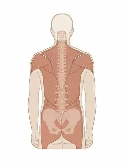
Background imageSpinal Collection: Back muscles, anatomical artwork

Back muscles, anatomical artwork
Back muscles. Anatomical computer artwork showing the muscles of the human back, and shoulders

Background imageSpinal Collection: Homo heidelbergensis vertebra C015 / 6799

Homo heidelbergensis vertebra C015 / 6799
Homo heidelbergensis vertebra. This fossil vertebra was excavated from the Sima de los Huesos pit in the Atapuerca foothills in Spain

Background imageSpinal Collection: Toothache, artwork C013 / 5927

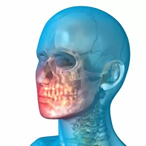
Toothache, artwork C013 / 5927
Toothache, computer artwork

Background imageSpinal Collection: Shoulder pain, artwork C013 / 5925

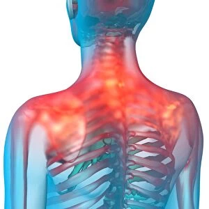
Shoulder pain, artwork C013 / 5925
Shoulder pain, computer artwork

Background imageSpinal Collection: Toothache, artwork C013 / 5926

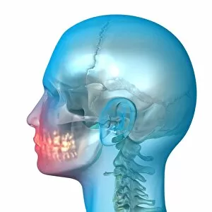
Toothache, artwork C013 / 5926
Toothache, computer artwork

Background imageSpinal Collection: Lower back pain, artwork C013 / 5922

Lower back pain, artwork C013 / 5922
Lower back pain, computer artwork

Background imageSpinal Collection: Neck pain, artwork C013 / 5923

Neck pain, artwork C013 / 5923
Neck pain, computer artwork

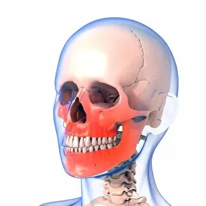
Background imageSpinal Collection: Jaw pain, artwork C013 / 5920

Jaw pain, artwork C013 / 5920
Jaw pain, computer artwork

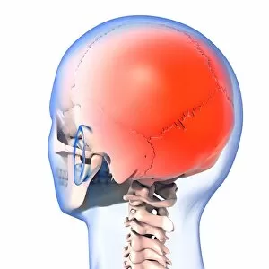
Background imageSpinal Collection: Headache, artwork C013 / 5916

Headache, artwork C013 / 5916
Headache, computer artwork

Background imageSpinal Collection: Headache, artwork C013 / 5915

Headache, artwork C013 / 5915
Headache, computer artwork

Background imageSpinal Collection: Headache, artwork C013 / 5914

Headache, artwork C013 / 5914
Headache, computer artwork

Background imageSpinal Collection: Headache, artwork C013 / 5913

Headache, artwork C013 / 5913
Headache, computer artwork

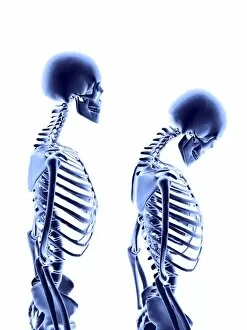
Background imageSpinal Collection: Osteoporosis C017 / 8250

Osteoporosis C017 / 8250
Osteoporosis. Computer artwork of a human female skeleton degenerating due to osteoporosis. At left is a normal skeleton. The degeneration, seen right

Background imageSpinal Collection: Osteoporosis C017 / 8248

Osteoporosis C017 / 8248
Osteoporosis. Computer artwork of a human female skeleton degenerating due to osteoporosis. At left is a normal skeleton. The degeneration, seen right
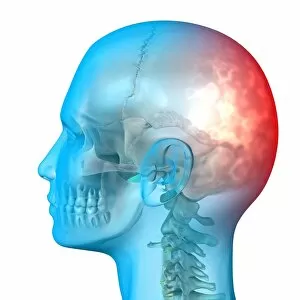
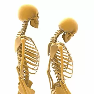
Background imageSpinal Collection: Osteoporosis C017 / 8245

Osteoporosis C017 / 8245
Osteoporosis. Computer artwork of a human female skeleton degenerating due to osteoporosis. At left is a normal skeleton. The degeneration, seen from left to right over time

Background imageSpinal Collection: Osteoporosis C017 / 8244

Osteoporosis C017 / 8244
Osteoporosis. Computer artwork of a human female skeleton degenerating due to osteoporosis. At left is a normal skeleton. The degeneration, seen right

Background imageSpinal Collection: Neck pain, artwork C013 / 8811

Neck pain, artwork C013 / 8811
Neck pain, computer artwork

Background imageSpinal Collection: Lower back pain, artwork C013 / 8810

Lower back pain, artwork C013 / 8810
Lower back pain, computer artwork

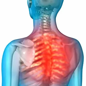
Background imageSpinal Collection: Back pain, artwork C013 / 7814

Back pain, artwork C013 / 7814
Back pain, computer artwork

Background imageSpinal Collection: Toothache, artwork C013 / 7813

Toothache, artwork C013 / 7813
Toothache, computer artwork

Background imageSpinal Collection: Toothache, artwork C013 / 7812

Toothache, artwork C013 / 7812
Toothache, computer artwork

Background imageSpinal Collection: Shoulder pain, artwork C013 / 7810

Shoulder pain, artwork C013 / 7810
Shoulder pain, computer artwork

Background imageSpinal Collection: Toothache, artwork C013 / 7811

Toothache, artwork C013 / 7811
Toothache, computer artwork

Background imageSpinal Collection: Headache, artwork C013 / 7797

Headache, artwork C013 / 7797
Headache, computer artwork

Background imageSpinal Collection: Headache, artwork C013 / 7796

Headache, artwork C013 / 7796
Headache, computer artwork

Background imageSpinal Collection: Headache, artwork C013 / 7795

Headache, artwork C013 / 7795
Headache, computer artwork

Background imageSpinal Collection: Coccyx pain, artwork C013 / 7793

Coccyx pain, artwork C013 / 7793
Coccyx (tailbone) pain, computer artwork

Background imageSpinal Collection: Chest muscles, artwork C013 / 4566

Chest muscles, artwork C013 / 4566
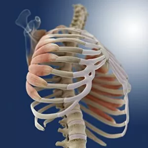
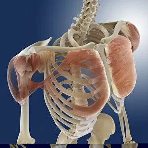
Chest muscles. Computer artwork of an oblique posterior view from above of some of the muscles (red) of the chest, showing their attachment to the bones of the ribcage and back

Background imageSpinal Collection: Chest muscles, artwork C013 / 4565

Chest muscles, artwork C013 / 4565
Chest muscles. Computer artwork of an oblique anterior view from below of some of the muscles (red) of the chest, showing their attachment to the bones of the ribcage and back

Background imageSpinal Collection: Chest muscles, artwork C013 / 4563

Chest muscles, artwork C013 / 4563
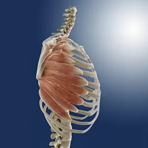
Chest muscles. Computer artwork of a side view of some of the muscles (red) of the chest, showing their attachment to the bones of the ribcage and back

Background imageSpinal Collection: Chest muscle, artwork C013 / 4561

Chest muscle, artwork C013 / 4561
Chest muscle. Computer artwork of an oblique anterior view of one of the muscles (red) of the chest and shoulders and its attachment to the bones of the back and ribcage

Background imageSpinal Collection: Shoulder muscles, artwork C013 / 4513

Shoulder muscles, artwork C013 / 4513
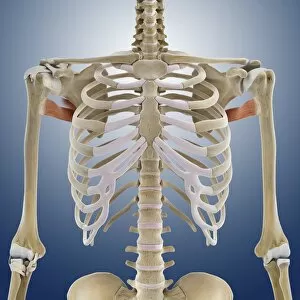
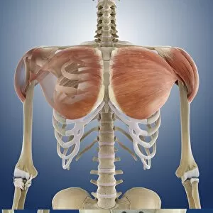
Shoulder muscles. Computer artwork of an anterior view of two of the muscles (red) of the shoulders and their attachment to the bones of the back and upper arms

Background imageSpinal Collection: Shoulder muscles, artwork C013 / 4515

Shoulder muscles, artwork C013 / 4515
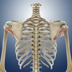
Shoulder muscles. Computer artwork of a posterior view of two of the muscles (red) of the shoulders and their attachment to the bones of the back and upper arms

Background imageSpinal Collection: Shoulder muscle, artwork C013 / 4508

Shoulder muscle, artwork C013 / 4508
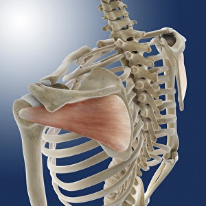
Shoulder muscle. Computer artwork of an oblique posterior view from below of one of the muscles (red) of the shoulders and its attachment to the bones of the back and upper arms

Background imageSpinal Collection: Shoulder muscles, artwork C013 / 4504

Shoulder muscles, artwork C013 / 4504
Shoulder muscles. Computer artwork of an oblique anterior view of two of the muscles (red) of the shoulders and their attachments to the bones of the chest, back and upper arms

Background imageSpinal Collection: Shoulder muscle, artwork C013 / 4507

Shoulder muscle, artwork C013 / 4507
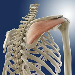
Shoulder muscle. Computer artwork of an oblique posterior view from above of one of the muscles (red) of the shoulders and its attachment to the bones of the back and upper arms

Background imageSpinal Collection: Shoulder muscles, artwork C013 / 4505

Shoulder muscles, artwork C013 / 4505
Shoulder muscles. Computer artwork of an anterior view of two of the muscles (red) of the shoulders and their attachments to the bones of the chest, back and upper arms


All Professionally Made to Order for Quick Shipping

"Unlocking the Secrets of Spinal Harmony: Exploring Chakras and the Nervous System" Delve into the intricate world of the spinal column, where ancient wisdom meets modern science. Discover how this remarkable structure, revered by pioneers like Ramon y Cajal, holds the key to our physical and spiritual well-being. Embark on a visual journey through time as you explore a diagram showcasing the human brain and spinal cord. Witness how these interconnected powerhouses orchestrate every aspect of our existence, from bodily functions to emotional experiences. Step back in time with Calots spinal surgery, an innovation from the 19th century that revolutionized medical practices. Marvel at illustrations depicting this groundbreaking procedure that aimed to alleviate pain and restore mobility. Peer into X-ray images capturing both normal necks and spines, revealing their intricate beauty hidden beneath our skin. Observe an extended neck vertebrae X-ray, shedding light on potential issues caused by misalignment or injury. Art comes alive as body pain is transformed into captivating artwork – a poignant reminder of the challenges faced by those afflicted with discomfort along their spine. Let your imagination soar as upper body skeletons take form through computer-generated artistry. Witness how lower back pain manifests itself in conceptual artwork – a vivid representation of its impact on daily life. Explore similar expressions for upper back pain; each stroke conveying tales of struggle yet resilience against this common ailment. Intriguingly juxtaposing these artistic interpretations are X-rays showcasing normal necks and spines - silent testaments to health and balance within our bodies' core structures. Uncover new perspectives on spinal health as you immerse yourself in this captivating collection.