Fraud Blocker Skip to main content

Scanning Electron Microscope Collection (page 2)

"Unveiling the Hidden World

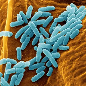
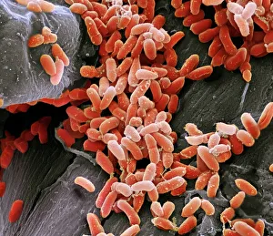
Background imageScanning Electron Microscope Collection: Pseudomonas aeruginosa bacteria, SEM

Pseudomonas aeruginosa bacteria, SEM
Pseudomonas aeruginosa bacteria, coloured scanning electron micrograph (SEM). These Gram-negative rod-shaped bacteria are found in soil and water, and as normal flora in the human intestine

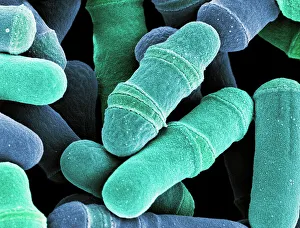
Background imageScanning Electron Microscope Collection: Dividing yeast cells, SEM

Dividing yeast cells, SEM
Dividing yeast cells. Coloured scanning electron micrograph (SEM) of Schizosaccharomyces pombe yeast cells dividing. S. pombe is a single-celled fungus that is studied widely as a model organism for

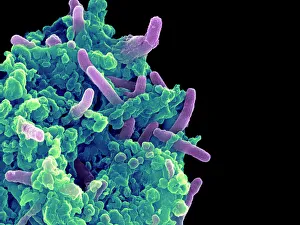
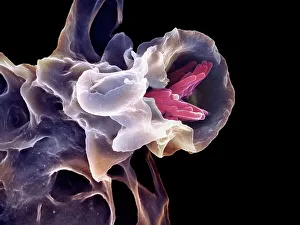
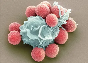
Background imageScanning Electron Microscope Collection: Bacteria infecting a macrophage, SEM

Bacteria infecting a macrophage, SEM
Bacteria infecting a macrophage. Coloured scanning electron micrograph (SEM) of Mycobacterium tuberculosis bacteria (purple) infecting a macrophage white blood cell

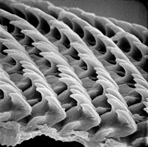
Background imageScanning Electron Microscope Collection: Snail teeth

Snail teeth

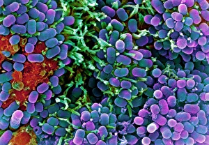
Background imageScanning Electron Microscope Collection: E. coli bacteria, SEM

E. coli bacteria, SEM
E. coli bacteria. Coloured scanning electron micrograph (SEM) of Escherichia coli bacteria (purple) taken from the small intestine of a child. E

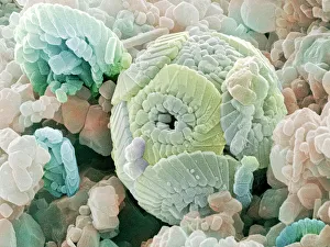
Background imageScanning Electron Microscope Collection: Calcareous phytoplankton fossil, SEM Z100 / 0213

Calcareous phytoplankton fossil, SEM Z100 / 0213
Calcareous phytoplankton fossil. Coloured scanning electron micrograph (SEM) of a fossil of the skeleton (coccosphere) of a coccolithotrope, a small marine algal organism

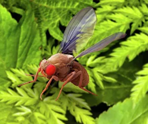
Background imageScanning Electron Microscope Collection: Fruit fly, SEM

Fruit fly, SEM
Fruit fly. Coloured scanning electron micrograph (SEM) of a male fruit fly (Drosophila melanogaster) flying past plant foliage. Its compound eyes (red) are seen and its wings are outstretched

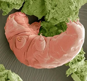
Background imageScanning Electron Microscope Collection: Water bear, SEM

Water bear, SEM
Water bear. Coloured scanning electron micrograph (SEM) of a water bear (Macrobiotus sp.), or tardigrade. This tiny invertebrate lives in aquatic and semi-aquatic habitats such as lichen

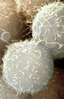
Background imageScanning Electron Microscope Collection: Stem cells, SEM

Stem cells, SEM
Stem cells, coloured scanning electron micrograph (SEM). Stem cells can differentiate into any other cell type. There are three main types of mammalian stem cell: embryonic stem cells

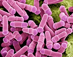
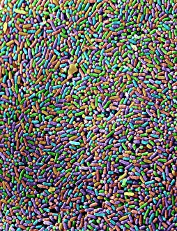
Background imageScanning Electron Microscope Collection: Salmonella bacteria, SEM

Salmonella bacteria, SEM
Salmonella bacteria, coloured scanning electron micrograph (SEM). Each rod-shaped structure is an individual bacterium. These bacteria occur mainly in human and animal intestines

Background imageScanning Electron Microscope Collection: Forget-me-not flower, SEM

Forget-me-not flower, SEM
Forget-me-not flower. Coloured scanning electron micrograph (SEM) of a forget-me-not (Myosotis arvensis) flower. The corolla consists of five petals (blue)

Background imageScanning Electron Microscope Collection: Pseudomonas aeruginosa bacteria, SEM

Pseudomonas aeruginosa bacteria, SEM
Pseudomonas aeruginosa bacteria, coloured scanning electron micrograph (SEM). These Gram-negative rod-shaped bacteria are found in soil and water, and as normal flora in the human intestine

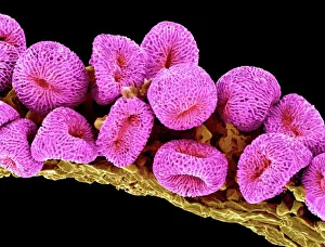
Background imageScanning Electron Microscope Collection: Geranium pollen, SEM

Geranium pollen, SEM
Geranium pollen. Coloured scanning electron micrograph (SEM) of pollen (pink) on the stamen of a geranium flower (Geranium sp.)

Background imageScanning Electron Microscope Collection: Plasmodium sp. malarial parasite

Plasmodium sp. malarial parasite
Scanning electron microscope image of a malarial protozoal parasite. The parasite requires the anopheles mosquito to complete its life cycle

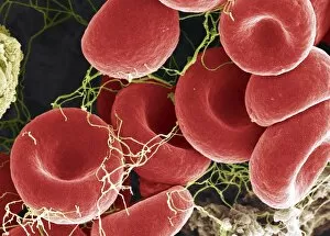
Background imageScanning Electron Microscope Collection: Blood clot, SEM

Blood clot, SEM
Blood clot, coloured scanning electron micrograph (SEM). Red blood cells are red and fibrin protein strands are green. Platelets are at bottom right

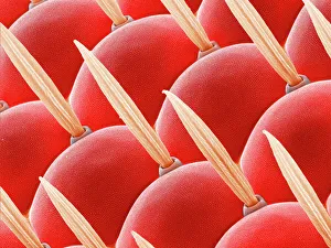
Background imageScanning Electron Microscope Collection: Compound eye of a fly, SEM Z340 / 0698

Compound eye of a fly, SEM Z340 / 0698
Compound eye. Coloured scanning electron micrograph (SEM) of the surface of a compound eye from a fruit fly (Drosophila busckii). The eye consists of many rounded lenses known as ommatidia

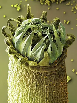
Background imageScanning Electron Microscope Collection: Moss capsule (Homalothecium sericeum) SEM

Moss capsule (Homalothecium sericeum) SEM
Moss spore capsule. Coloured scanning electron micrograph (SEM) of part of the open mouth of a capsule (spore case) of a the moss Homalothecium sericeum

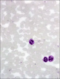
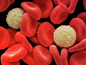
Background imageScanning Electron Microscope Collection: Red and white blood cells, SEM

Red and white blood cells, SEM
Human red and white blood cells, coloured scanning electron micrograph (SEM). Magnification x5167 at an image size of 10 cm wide

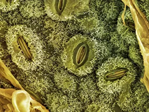
Background imageScanning Electron Microscope Collection: English oak leaf pores, SEM

English oak leaf pores, SEM
English oak leaf pores. Coloured scanning electron micrograph (SEM) of stomata (round) on the underside of a leaf from an English oak (Quercus robur) tree

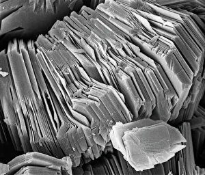
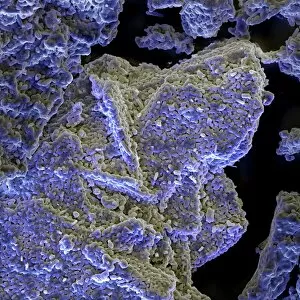
Background imageScanning Electron Microscope Collection: Kaolinite

Kaolinite
Scanning electron microscope image of kaolinite (x 4.00K). Its a common phyllosilicate mineral, its structure is composed of silicate sheets bonded to aluminum oxide/hydroxide layers

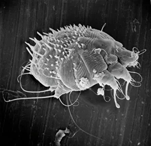
Background imageScanning Electron Microscope Collection: Sarcoptes scabiei, scabies mite

Sarcoptes scabiei, scabies mite
Scanning electron microscope image of an itch or scabies mite, a parasite that infests a wide variety of mammalian hosts including humans

Background imageScanning Electron Microscope Collection: Tendon, SEM

Tendon, SEM
Tendon, coloured scanning electron micrograph (SEM), showing bundles of collagen fibres. Magnification: x5000 when printed at 10 centimetres wide

Background imageScanning Electron Microscope Collection: Macrophage engulfing TB bacteria, SEM

Macrophage engulfing TB bacteria, SEM
Macrophage engulfing TB bacteria. Coloured scanning electron micrograph (SEM) of a macrophage white blood cell (purple) engulfing a tuberculosis (Mycobacterium tuberculosis) bacterium (pink)

Background imageScanning Electron Microscope Collection: Scanning Electron Micrograph (SEM): Crab Spider, Magnification x 140 (A4 size: 29. 7 cm width)

Scanning Electron Micrograph (SEM): Crab Spider, Magnification x 140 (A4 size: 29. 7 cm width)
LRDS-120 Crab Spider Scanning Electron Micrograph (SEM) Xysticus sp. Magnification x 140 (A4 size: 29.7 cm width) Coloured by hand to enhance natural features

Background imageScanning Electron Microscope Collection: Scanning Electron Micrograph (SEM): House Fly, Magnification x 40 (A4 size: 29. 7 cm width)

Scanning Electron Micrograph (SEM): House Fly, Magnification x 40 (A4 size: 29. 7 cm width)
LRDS-72 House Fly Scanning Electron Micrograph (SEM) Musca domestica Magnification x 40 (A4 size: 29.7 cm width) Coloured by hand to enhance natural features

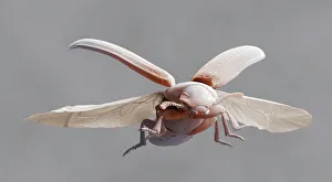
Background imageScanning Electron Microscope Collection: Red flour beetle in flight

Red flour beetle in flight
Red flour beetle. Coloured scanning electron micrograph of a red flour beetle (Tribolium castaneum) in flight. One of the most significant pests of dried foods and the milling industry

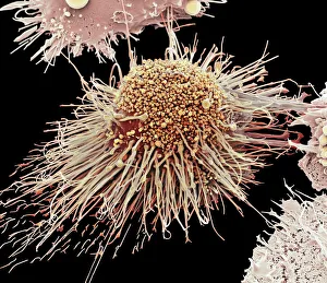
Background imageScanning Electron Microscope Collection: Activated macrophage, SEM C015 / 6375

Activated macrophage, SEM C015 / 6375
Activated macrophage. Coloured scanning electron micrograph (SEM) of an active macrophage white blood cell. Macrophages are cells of the bodys immune system that are found in the tissues rather than

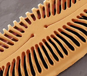
Background imageScanning Electron Microscope Collection: Diatom frustule, SEM

Diatom frustule, SEM
Diatom frustule. Coloured scanning electron micrograph (SEM) of the frustule of a Pinnularia sp. diatom. The diatoms are a group of photosynthetic, single-celled algae containing about 10

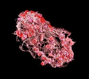
Background imageScanning Electron Microscope Collection: Blood clot, SEM C016 / 9747

Blood clot, SEM C016 / 9747
Blood clot, coloured scanning electron micrograph (SEM). Red blood cells (erythrocytes) are trapped within a fibrin protein mesh (beige)

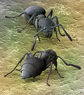
Background imageScanning Electron Microscope Collection: Tropical ants, SEM

Tropical ants, SEM
Tropical ants. Coloured scanning electron micrograph (SEM) of two small, tropical ants (family Formicidae, 2 millimetres long)

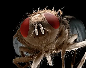
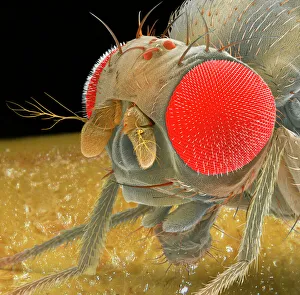
Background imageScanning Electron Microscope Collection: Fruit fly, SEM Z340 / 0699

Fruit fly, SEM Z340 / 0699
Fruit fly. Coloured scanning electron micrograph (SEM) of the head of a fruit fly (Drosophila busckii). Its two compound eyes (red) are seen on either side of the head

Background imageScanning Electron Microscope Collection: Stalk-eyed fly, SEM C014 / 4882

Stalk-eyed fly, SEM C014 / 4882
Stalk-eyed fly. Coloured scanning electron micrograph (SEM) of the head of a stalk-eyed fly (family Diopsidae). Stalk-eyed flies are mostly tropical insects

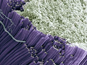
Background imageScanning Electron Microscope Collection: Rod and cone cells of the eye, SEM C014 / 4866

Rod and cone cells of the eye, SEM C014 / 4866
Rod and cone cells of the eye. Coloured scanning electron micrograph (SEM) of rod and cone cells in the retina of a mammalian eye

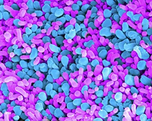
Background imageScanning Electron Microscope Collection: Baking soda crystals, SEM C016 / 8041

Baking soda crystals, SEM C016 / 8041
Sodium bicarbonate. Coloured scanning electron micrograph (SEM) of crystals of sodium bicarbonate (or sodium hydrogen carbonate). Sodium bicarbonate is a white solid commonly used in baking powder

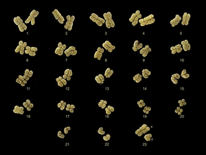
Background imageScanning Electron Microscope Collection: Full set of male chromosomes, SEM

Full set of male chromosomes, SEM
Set of human chromosomes, coloured scanning election micrograph (SEM). Chromosomes are a packaged form of the genetic material deoxyribonucleic acid (DNA)

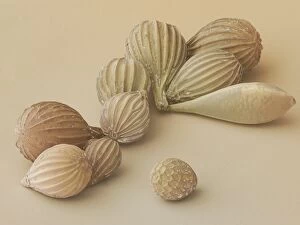
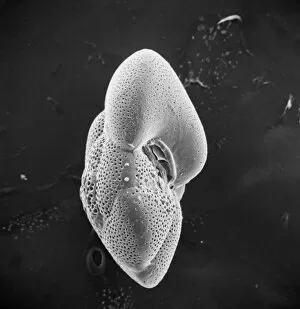
Background imageScanning Electron Microscope Collection: Foraminifera, SEM

Foraminifera, SEM
Foraminifera. Coloured scanning electron micrograph (SEM) of a selection of foraminiferans, including Oolina hexagona (bottom right) and a number of Lagena sp

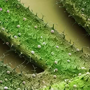
Background imageScanning Electron Microscope Collection: French lavender leaf surface, SEM

French lavender leaf surface, SEM
French lavender leaf surface. Coloured scanning electron micrograph (SEM) of a section of French lavender (Lavandula dentata) leaf, showing the tooth-like structures (trichomes)

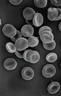
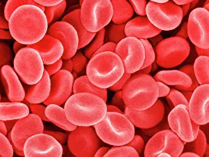
Background imageScanning Electron Microscope Collection: Human red blood corpuscles

Human red blood corpuscles
Scanning electron microscope (SEM) of red blood cells showing their characteristic biconcave shape which increases the surface area for diffusion

Background imageScanning Electron Microscope Collection: Globorotalia scitula, foraminifera fossil

Globorotalia scitula, foraminifera fossil
Scanning electron microscope (SEM) image showing a fossilised planktonic species of foraminifera

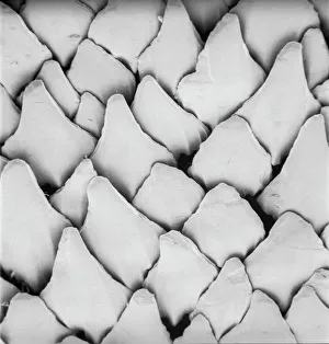
Background imageScanning Electron Microscope Collection: Scyliorhinus canicula, dogfish

Scyliorhinus canicula, dogfish
Scanning electron microscope (SEM) image of the scales of a dogfish (x 40)

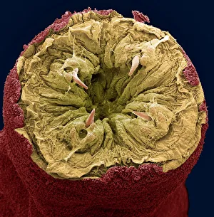
Background imageScanning Electron Microscope Collection: Ragworm mouth, SEM

Ragworm mouth, SEM
Ragworm mouth. Coloured scanning electron micrograph (SEM) of the mouth of a ragworm (Nereis sp.). Four teeth (cream) are seen around the opening. These worms are used commercially as fishing bait

Background imageScanning Electron Microscope Collection: Head of a young newt, SEM

Head of a young newt, SEM
Head of a young newt. Scanning electron micrograph (SEM) of a juvenile (immature) newt. Although this newt is in the last stages of development

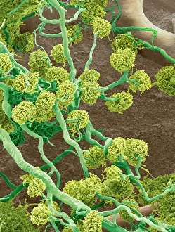
Background imageScanning Electron Microscope Collection: Kidney glomeruli, SEM

Kidney glomeruli, SEM
Kidney glomeruli, coloured scanning electron micrograph (SEM). The cells that usually surround the glomeruli have been removed to reveal the coiled capillaries at the core of the glomeruli

Background imageScanning Electron Microscope Collection: Red blood cells, SEM

Red blood cells, SEM
Red blood cells, coloured scanning electron micrograph (SEM). Red blood cells (erythrocytes) are biconcave, giving them a large surface area for gas exchange, and highly elastic

Background imageScanning Electron Microscope Collection: Phagocytosis of fungal spores, SEM

Phagocytosis of fungal spores, SEM
Phagocytosis of a fungus spore. Coloured scanning electron micrograph (SEM) of a neutrophil (blue) phagocytosing (engulfing and destroying) spores from the fungus Aspergillus fumigatus (red)

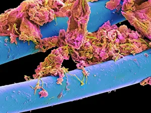
Background imageScanning Electron Microscope Collection: Used dental floss, SEM

Used dental floss, SEM
Used dental floss. Coloured scanning electron micrograph (SEM) of dental plaque on strands of used dental floss. Plaque consists of a film of bacteria embedded in a glycoprotein matrix

Background imageScanning Electron Microscope Collection: Neutrophil cell trapping bacteria, SEM

Neutrophil cell trapping bacteria, SEM
Neutrophil cell trapping bacteria. Coloured scanning electron micrograph (SEM) of bacteria (rod-shaped) being trapped by a neutrophil cell


All Professionally Made to Order for Quick Shipping

"Unveiling the Hidden World: Exploring with a Scanning Electron Microscope" The scanning electron microscope (SEM) is an incredible tool that allows us to delve into the microscopic realm, revealing intricate details and unlocking secrets of the unseen. With its high magnification capabilities, it offers a glimpse into fascinating aspects of life and nature. In one captivating image, we witness the beauty of gender determination as X and Y chromosomes are captured under the SEM's lens. The intricate patterns and structures within these chromosomes showcase the complexity of our genetic makeup. Moving on to another astonishing discovery, a Scanning Electron Micrograph presents us with an up-close view of a Praying Mantis at 30 times magnification. Every tiny detail becomes visible, from its delicate limbs to its mesmerizing eyes – reminding us of nature's remarkable diversity. Shifting focus to plant life, lavender pollen grains come alive in another SEM image. These minuscule particles take center stage as their unique shapes and textures become apparent under intense scrutiny. Zooming in even further reveals a Tardigrade or 'Water Bear' at an astounding 1250 times magnification. This resilient creature captures our imagination as we explore its alien-like features through this powerful imaging technique. Delving deeper into biological wonders, T lymphocytes battling cancer cells are showcased in yet another breathtaking SEM capture. This visual representation highlights the ongoing fight within our bodies against diseases like cancer – emphasizing both hope and resilience. Calcareous phytoplankton takes center stage next as we uncover their intricate forms through SEM imagery. These microscopic organisms play crucial roles in marine ecosystems while leaving us awe-inspired by their stunningly detailed structures. Transitioning to neural marvels, nerve cells reveal their intricacy under the watchful eye of an SEM. Their branching extensions create complex networks that enable communication throughout our bodies – showcasing nature's ingenuity at work. Venturing into insect realms once again, a fruit fly is immortalized under the SEM's lens.